© Frederik Köpper / Max Planck Institute for Biophysical Chemistry
News • Downsized imaging
Signals from a miniature MRI unit
Magnetic resonance imaging (MRI) is indispensable in medical diagnostics. However, MRI units are large and expensive to acquire and operate. With smaller and cost-efficient systems, MRI would be more flexible and more people could benefit from the technique.
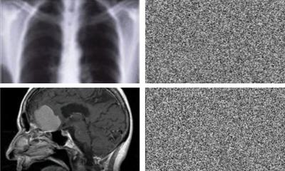
Such miniature MRI units generate a much weaker signal that is difficult to analyze, though. Researchers at the Göttingen Max Planck Institute (MPI) for Biophysical Chemistry and the Center for Biostructural Imaging of Neurodegeneration of the University Medical Center Göttingen, Germany, have now developed a method amplifying the signal so much that they could monitor a metabolic reaction in real time with a miniature MRI. This is an important contribution to making flexible small MRI devices usable.
The researchers now published their findings in the journal Chemical Science.
That was an exciting technical fiddle. We had to coil more than a kilometer of copper wire
Stefan Glöggler
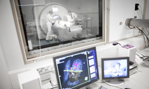
MRI provides razor-sharp images of our inner body and is used to diagnose a wide range of diseases ranging from inflammation to arteriosclerosis and cancer. However, conventional MRI units are huge, filling entire rooms. The impressive size has its reason: The donut-shaped tube contains large magnets that generate a very strong magnetic field. When combined with short radio frequency pulses, the magnetic force elicits a signal from the water in the patient’s body. The signal gives rise to an image of the body region examined. The stronger the magnetic field, the clearer the signal – and hence the more detailed the image produced.
The power of these MRI units, however, brings along two decisive drawbacks: Firstly, because of their size and weight, the machines are not mobile and cannot be brought directly to the patient’s bed. Secondly, their operation is very costly: It requires a lot of electricity and expensive liquids such as helium at minus 270 degrees Celsius to cool the magnets. For these reasons, only financially strong institutions can run such MRI units. Most people worldwide still do not have access to this technology.
© Frederik Köpper / Max Planck Institute for Biophysical Chemistry
Several scientists want to change this and are developing inexpensive, mobile MRI devices. One of them is Stefan Glöggler, research group leader at the Göttingen MPI for Biophysical Chemistry and at the Center for Biostructural Imaging of Neurodegeneration. “A major technical challenge in the design of a miniature MRI unit is that the signal generated is very weak,” Glöggler explains. “This is because such a small device has to operate with much weaker magnets than a conventional system. We have now succeeded in significantly amplifying the signal.”
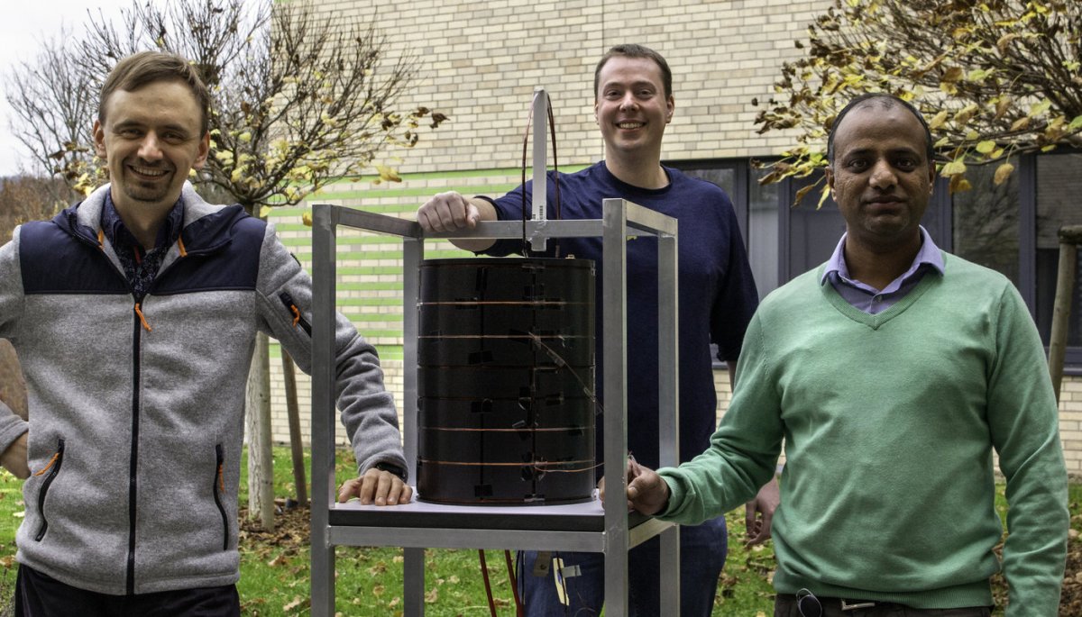
For their experiments, the Göttingen chemists have constructed their own miniature MRI unit. It is about the size of a small barrel. “That was an exciting technical fiddle. We had to coil more than a kilometer of copper wire,” Glöggler says. “We had great support from the precision mechanics, electronics engineers, and carpenters at our Max Planck Institute. We would not have been able to realize the project without them.”
The magnetic field is about a hundred times lower than in conventional MRI devices. Its strength is comparable to that of magnets we attach to the fridge at home
Sergey Korchak
“Our little tomograph is very flexible. It can be adapted to the size of the object to be examined – be it a chemical sample or a human head,” reports Sergey Korchak, a postdoctoral researcher in Glöggler’s team. “The magnetic field is about a hundred times lower than in conventional MRI devices. Its strength is comparable to that of magnets we attach to the fridge at home.”
The scientists now transferred a method already established in conventional MRI units, the so-called hyperpolarization, to their low-field device. This enabled them to amplify the signal at the weak magnetic field so much that it became measurable. With this improvement, they could follow in real time how pyruvate is converted into lactate by using a miniature MRI for the first time. The biochemical reaction continuously takes place in our body cells as part of the energy production process and it was not chosen at random by the Göttingen researchers, as Glöggler's postdoc Anil Jagtap says: “How much pyruvate cells convert into lactate provides information about whether sufficient oxygen is available in a tissue or whether it is used to produce energy. In the future, this information could help to diagnose brain traumas and certain types of cancer.” Corresponding MRI studies are already underway in hospitals.
Glöggler is optimistic that such examinations will soon be possible with low-field MRI devices. “The signal amplification we have developed is an important piece of the puzzle for developing portable MRI units to market maturity, so that more patients can benefit from the diagnostic strength of this technology,” the chemist emphasizes.
Source: Max Planck Institute (MPI) for Biophysical Chemistry
24.11.2020