Image source: Cribaro et al, Acta Neuropathologica Communications 2021 (CC-BY 4.0)
News • Brain tumour analysis
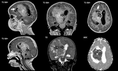
Glioblastoma '3D maps' help find new therapies
Researchers at the Universitat Autònoma de Barcelona obtained a highly accurate recreation of human glioblastoma’s features using a novel 3D microscopy analysis.
The study, published in the journal Acta Neuropathologica Communications, provides new information to help with the diagnose, by finding therapeutical targets and designing immunotherapeutical strategies.
This local and populational differentiation [of immune cells] could be an important factor that may help diagnosis and aid in the search for new therapeutic targets
Carlos Barcia
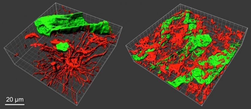
This new analysis of 3D images and quantitative data “will help to appreciate from within how the tumor is built in its full dimensionality, and to identify where different cell types are located”, explains George Paul Cribaro, first author of the study. “It provides more complete information than the usual 2D analyses performed for neuropathological diagnosis”
With this new approach, authors show the alterations in tumor blood vessels, and that these vascular wall abnormalities do not hinder the entrance of lymphocytes T (potential defense against tumoral cells), which is relevant for the design and use of immunotherapies targeting malignant cells. Moreover, the images allow the tumor to be differentiated into two areas, the tumor tissue properly speaking, and the stroma, which gives support to the tumor, in which there are different immunological microenvironments. “Immune cells like microglia and macrophages are seen in both areas, but they are shaped by different subpopulations. This local and populational differentiation could be an important factor that may help diagnosis and aid in the search for new therapeutic targets”, indicates Carlos Barcia, coordinator of the work.
The work provides a set of resource images that will facilitate the understanding of the complexity of this tumor, showing some aspects to be considered when designing new therapeutic approaches.
Source: Universitat Autònoma de Barcelona
22.02.2021