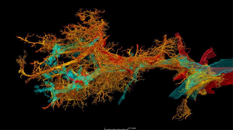
Image source: Ackermann et al., eBioMedicine 2022 (CC BY 4.0)
News • New X-ray technique
HiP-CT reveals link between long Covid and pulmonary fibrosis
A new X-ray technology co-developed by University College London (UCL) has been used to identify a link between the damage that severe Covid-19 can inflict on lungs and pulmonary fibrosis, a disease that causes severe scarring of lung tissue.
The high-energy X-ray technique, Hierarchical Phase-Contrast Tomography (HiP-CT), scans whole organs down to cellular level, allowing clinicians to view blood vessels about a tenth of the diameter of a human hair. Experts have now used the technology to reveal microscopic lung clots and changes in blood vessels that have only been found in pulmonary fibrosis associated with Covid-19, further improving our understanding of the long-term effects of serious Covid-19 infection. Around 20% of patients who survive severe Covid-19 (requiring hospitalisation) go on to develop pulmonary fibrosis. Typical life expectancy for the disease is around three to five years after diagnosis.
For the paper, published in eBioMedicine, experts examined the intact lungs of patients who had passed away after contracting Covid-19. To do this they used HiP-CT, a particle accelerator that provides the world’s brightest X-ray source, at around 100 billion times brighter than a hospital X-ray, developed by UCL with scientists from the European Synchrotron Research Facility (ESRF).
Recommended article
News • Coronavirus imaging
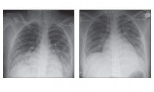
Brightest ever X-ray shows lung damage from Covid-19
The damage caused by Covid-19 to the lungs’ smallest blood vessels has been intricately captured using high-energy X-rays emitted by a special type of particle accelerator. Scientists used a new imaging technology called Hierarchical Phase-Contrast Tomography (HiP-CT), to scan donated human organs, including lungs from a Covid-19 donor.
The team, from RWTH Aachen University Hospital, Hannover Medical School, HELIOS University Hospital in Wuppertal and the University Medical Centre Mainz, all in Germany, together with UCL and the ESRF, discovered a distinctive mosaic-like pattern of damage in the lungs that hasn’t been seen before. The experts also compared blood samples from Covid-19 patients with those from some common and serious lung diseases including pulmonary fibrosis unrelated to Covid-19. The team conclude that the microscopic clots, seen within tiny blood vessels, cause the distinctive pattern and drive inflammation in the lungs that can lead to long Covid symptoms such as shortness of breath and disease such as pulmonary fibrosis. The team hope that identifying the tiny clots found in damaged lung tissue and the changes in microscopic blood vessels can serve as an early indicator of Covid-induced pulmonary fibrosis, for which there is no cure, and allow clinicians time to administer treatment to potentially lessen the impact and improve patients’ quality of life.
With the new technology of HiP-CT, we were able to see, for the first time, that the scarring processes in post-Covid fibrosis are the result of generalised vascular damage caused by the SARS-CoV-2 virus
Danny Jonigk
Co-author Dr Claire Walsh (UCL Mechanical Engineering), who co-developed HiP-CT with ESRF, said: “A technique like HiP-CT is a real game-changer for unveiling not only the really fine detail of the tissue damage, but also how it is distributed across a whole organ and relating that back to what is seen in clinic. This technology allows us to understand more about the impact that severe Covid-19 can have on the lungs and will help us when diagnosing and treating pulmonary fibrosis resulting from the virus.”
Co-author Professor Danny Jonigk (RWTH Aachen University Hospital) said: “These heterogeneously distributed, distinct changes at the level of the finest lung lobules cannot be detected via clinical imaging due to the lack of detail in current clinical technology. With the new technology of HiP-CT, we were able to see, for the first time, that the scarring processes in post-Covid fibrosis are the result of generalised vascular damage caused by the SARS-CoV-2 virus.” The authors say that treatments such as giving oxygen at an earlier stage of severe Covid-19 might be beneficial for patients with a severe form of the disease in reducing scarring in the lungs.
HiP-CT was developed jointly by UCL and ESRF, a project led by Professor Peter Lee (UCL Mechanical Engineering). He said: “We’re continuing to develop HiP-CT with ESRF to better understand the effects of other diseases on our organs, including the heart and brain, hopefully providing new insights into conditions such as congenital heart disease and Alzheimer’s. HiP-CT is a real breakthrough as it obtains near cellular resolution over entire organs.”
Source: University College London
09.11.2022