News • Key findings in perivascular spaces
Ultra-high-res MRI reveals how migraine changes the brain
For the first time, a new study has identified enlarged perivascular spaces in the brains of migraine sufferers. Results of the study will be presented at the annual meeting of the Radiological Society of North America (RSNA).
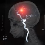
Image source: RSNA
"In people with chronic migraine and episodic migraine without aura, there are significant changes in the perivascular spaces of a brain region called the centrum semiovale," said study co-author Wilson Xu, an M.D. candidate at Keck School of Medicine of the University of Southern California in Los Angeles. "These changes have never been reported before."
Migraine is a common, often debilitating condition, involving a severe recurring headache. Migraines may also cause nausea, weakness and light sensitivity. According to the American Migraine Foundation, over 37 million people in the U.S. are affected by migraine, and up to 148 million people worldwide suffer from chronic migraine. Perivascular spaces are fluid-filled spaces surrounding blood vessels in the brain. They are most commonly located in the basal ganglia and white matter of the cerebrum, and along the optic tract. Perivascular spaces are affected by several factors, including abnormalities at the blood-brain barrier and inflammation. Enlarged perivascular spaces can be a signal of underlying small vessel disease. "Perivascular spaces are part of a fluid clearance system in the brain," Xu said. "Studying how they contribute to migraine could help us better understand the complexities of how migraines occur."
Xu and colleagues set out to determine the association between migraine and enlarged perivascular spaces. The researchers used ultra-high-field 7T MRI to compare structural microvascular changes in different types of migraine. "To our knowledge, this is first study using ultra-high-resolution MRI to study microvascular changes in the brain due to migraine, particularly in perivascular spaces," Xu said. "Because 7T MRI is able to create images of the brain with much higher resolution and better quality than other MRI types, it can be used to demonstrate much smaller changes that happen in brain tissue after a migraine."
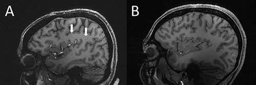
Image source: RSNA
Study participants included 10 with chronic migraine, 10 with episodic migraine without aura, and five age-matched healthy controls. All patients were between 25 and 60 years old. Patients with overt cognitive impairment, brain tumor, prior intracranial surgery, MRI contraindications and claustrophobia were excluded from the study. The researchers calculated enlarged perivascular spaces in the centrum semiovale (central area of white matter) and basal ganglia areas of the brain. White matter hyperintensities—lesions that "light up" on MRI—were measured using the Fazekas scale. Cerebral microbleeds were rated with the microbleed anatomical rating scale. The researchers also collected clinical data such as disease duration and severity, symptoms at time of scan, presence of aura and side of headache.
Recommended article
Article • Brain imaging
White matter hyperintensities: a valuable biomarker to assess mortality risk
White matter hyperintensities (WMH) on the brain seen on MRI represent a biomarker associated with a 50/50 risk of death within five years after a first incident acute ischemic stroke (AIS) or transient ischemic attack (TIA).
Statistical analysis revealed that the number of enlarged perivascular spaces in the centrum semiovale was significantly higher in patients with migraine compared to healthy controls. In addition, enlarged perivascular space quantity in the centrum semiovale correlated with deep white matter hyperintensity severity in migraine patients. "We studied chronic migraine and episodic migraine without aura and found that, for both types of migraine, perivascular spaces were bigger in the centrum semiovale," Xu said. "Although we didn't find any significant changes in the severity of white matter lesions in patients with and without migraine, these white matter lesions were significantly linked to the presence of enlarged perivascular spaces. This suggests that changes in perivascular spaces could lead to future development of more white matter lesions."

Image source: RSNA
The researchers hypothesize that significant differences in the perivascular spaces in patients with migraine compared to the healthy controls might be suggestive of glymphatic disruption within the brain. The glymphatic system is a waste clearance system that utilizes perivascular channels to help eliminate soluble proteins and metabolites from the central nervous system. However, whether such changes affect migraine development or result from migraine is unknown.
Continued study with larger case populations and longitudinal follow-up will better establish the relationship between structural changes and migraine development and type. "The results of our study could help inspire future, larger-scale studies to continue investigating how changes in the brain's microscopic vessels and blood supply contribute to different migraine types," Xu said. "Eventually, this could help us develop new, personalized ways to diagnose and treat migraine."
Source: Radiological Society of North America
25.11.2022