Image source: University of Notre Dame
News • From science fiction to reality
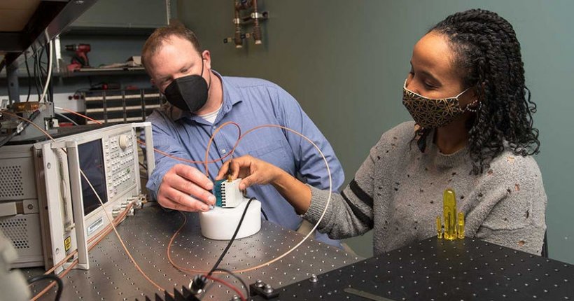
Researchers develop powerful pocket-sized imaging device
Before Wilhelm Röntgen, a mechanical engineer, discovered a new type of electromagnetic radiation in 1895, physicians could only dream of being able to see inside the body. Within a year of Röntgen’s discovery, X-rays were being used to identify tumors. Within 10 years, hospitals were using X-rays to help diagnose and treat patients.
In 1972, computed tomography (CT) scans were developed. In the 1980s, magnetic resonance imaging (MRI) technology became commercially available. Today, engineers like Thomas O’Sullivan, assistant professor of electrical engineering at the University of Notre Dame, continue the quest to improve the quality of medical diagnosis and treatment using near-infrared optical imaging. O’Sullivan and his team are developing a powerful, pocket-sized optical imager that may once have seemed like the stuff of science fiction.
“When envisioning medicine of the future, many people think of Dr. Leonard McCoy, a physician in the original 'Star Trek’ series,” said O’Sullivan. “Dr. McCoy used a handheld tricorder to scan an individual and immediately assess injuries or diseases anywhere in the body. We don’t have a commercial product like McCoy’s tricorder yet, but we’re close to making it a reality. We’re developing new medical imaging technology that uses light to give us a better view of the function of tissues and cells deep under the skin in a way that is safe and relatively low-cost. Equally important, we’re scaling that technology down to fit in a doctor’s pocket so it is as portable as a stethoscope or a thermometer.”
Recommended article

News • Genome sequence analyzer app
Researchers build world’s first DNA “tricorder”
Cold Spring Harbor Laboratory (CSHL) scientists developed the world’s first mobile genome sequence analyzer, a new iPhone app called iGenomics. By pairing an iPhone with a handheld DNA sequencer, users can create a mobile genetics laboratory, reminiscent of the “tricorder” featured in Star Trek.
CT and MRI scans revolutionized the way diseases were diagnosed and monitored, O’Sullivan said. But they can’t be used frequently. CT scans use low doses of radiation; a single scan takes about 10 minutes and costs up to $2,500. MRI uses magnetic and radio waves. One scan takes up to an hour and can cost as much as $4,000. Both procedures require entire rooms of hardware. “The small device we’re developing uses light, which is very safe, to help a doctor scan for the earliest signs of disease during an office visit — or even give you a device to take home,” O’Sullivan said. “The device also could be used to monitor progression and treatment — from breast cancer to brain function to personal health — with more exacting precision.”
The new platform collects large amounts of data quickly, reconstructing it into 2D and 3D images, O’Sullivan said.
Source: University of Notre Dame
19.02.2021