© beerkoff - stock.adobe.com
News • Tissue analysis tool
'Smart scalpel' to help remove brain tumours
When a surgeon is removing a tumour, it’s not always easy to make sure that all the cancer cells have been eradicated.
Removing too much healthy tissue is obviously undesirable, and sparing organ tissue is vital, especially when it comes to the brain. In recent years, scientists at the Maastricht MultiModal Molecular Institute (M4I) have therefore developed an ‘intelligent knife’, or iKnife. The European subsidy programme Interreg Flanders-Netherlands has made more than two million euros available for the further development of this technology. In partnership with UZ Leuven, the Maastricht scientists have now completed an extensive study on the instrument’s clinical application.
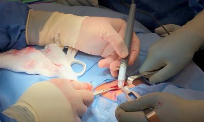
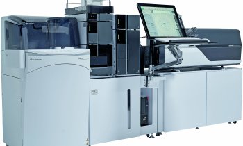
This showed that the iKnife is able to recognise tissue from brain tumour in seconds as it cuts, with more than 98% accuracy. The iKnife is an electrosurgical instrument that creates smoke when it comes into contact with tissue during the operation. The smoke contains molecules that are a kind of fingerprint of the tissue. This provides information beyond what is visible, enabling the surgeon to distinguish a tumour from surrounding tissue. During previous tests, however, the large analysis device proved to be a major obstacle in the operating theatre. The research team is therefore now working on a handy screener that is much more user-friendly for brain surgeons.
Recommended article

Article • Mass spectrometry goes handheld
A pen to pin down the fringes of cancer
Mass spectrometry – a powerful tool for analysing the molecular composition of a tissue sample – is invaluable during cancer surgery. However, mass spectrometers are complex and unwieldy, and certainly a poor fit for an operating room (OR). To create a bridge between the lab and OR, Professor Livia S Eberlin, from Baylor College of Medicine, has developed a very special ‘pen’.
‘Making our current iKnife, or molecular navigator, more compact is difficult, because as much as possible we want to avoid affecting the sensitivity and the accuracy of the present system,’ says M4I researcher Eva Cuypers. ‘This means we are facing some tough technical challenges. For example, we have to use a different type of detector to make everything compact. As a result, we will also have to develop a more compact component for the smoke input. Ultimately, the principle remains the same, so we can compare the molecular profiles provided by the new system with those of our previously developed model. Artificial intelligence will help us to see whether it’s really possible for us to translate our current model to the new system.’
Once we have sufficiently proven that the technique offers benefits for this specific patient group, it’s even conceivable that we will use it to treat other types of patients, such as those with severe epilepsy
Olaf Schijns
The iKnife is based on the analysis technique of mass spectrometry. It enables a significant improvement in surgical decisions. Around 60% of brain tumour patients experience a relapse within five years of surgery, because the tumour has grown back from residual cells. What can the more precise tissue recognition offered by the iKnife mean in practice for individual brain tumour patients?
‘The iKnife will most likely lead to faster recognition of tumour cells during brain surgery,’ says Maastricht neurosurgeon Olaf Schijns. ‘This is possible thanks to the analysis of molecules in the smoke produced when using the iKnife. It allows us to remove a brain tumour faster and more completely, without affecting healthy brain tissue. Once we have sufficiently proven that the technique offers benefits for this specific patient group, it’s even conceivable that we will use it to treat other types of patients, such as those with severe epilepsy.’
Source: Maastricht University
18.03.2024


