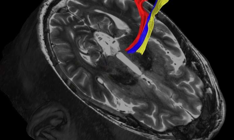
Credit: UT Southwestern Medical Center
News • Neuro-Imaging
Advanced MRI scans may improve treatment of Parkinson's disease
Recently developed MRI techniques used to more precisely target a small area in the brain linked to Parkinson's disease and essential tremor may lead to better outcomes without surgery and with less risk of negative effects, a new study led by UT Southwestern researchers suggests.
The study describes recently refined MRI methods designed to allow neuroradiologists to zero in on a pea-sized region in the brain's thalamus involved in movement. Using the images, doctors then can use high-intensity focused ultrasound (HIFU) to ablate, or burn away, problem tissue, says Bhavya R. Shah, M.D., first author of the study and an assistant professor of radiology and neurological surgery at UT Southwestern's Peter O'Donnell Jr. Brain Institute. "The benefit for patients is that we will be better able to target the brain structures that we want," Shah says. "And because we're not hitting the wrong target, we'll have fewer adverse effects." The procedures are already Food and Drug Administration-approved for use in patients, and UTSW plans to begin employing them to treat patients when its Neuro High Intensity Focused Ultrasound Program opens this fall.
Adverse effects from imprecise targeting include problems walking or slurring words. While such effects are usually temporary, they can be permanent in 15 to 20 percent of cases, says Dr. Shah.
According to the National Institutes of Health, essential tremor affects up to 10 million Americans and Parkinson's disease impacts more than 1 million. Both are neurologic diseases thought to have genetic links. The first line of treatment for the involuntary trembling or shaking seen with these diseases is medication. However, approximately 30 percent of patients do not respond well to drugs, according to the study.
In the late 1990s, neurosurgeons began using a procedure called deep brain stimulation, opening the skull to permanently implant metal electrodes that could then be stimulated via a battery pack. About a decade ago, a new MRI-guided procedure emerged that uses high-intensity ultrasound waves to heat and eliminate a small section of the thalamus linked to the disorders. MRI-guided HIFU is currently approved for treatment of essential tremor and tremors seen in Parkinson's disease patients. The outpatient procedure does not require opening the skull, and the patient is awake while it is performed, says Dr. Shah. "No cuts. No anesthesia. No implanted devices."
A challenge in both procedures has been locating the precise area inside the brain's thalamus to treat—the pea-sized ventral intermediate nucleus, says Dr. Shah. Traditionally, doctors have relied on either landmarks or maps of the brain drawn from cadavers to help them pinpoint the correct location. However, every brain is different, Dr. Shah says, and tiny errors can lead to damage in surrounding tissue, or to missing portions of the correct target.
Three newly refined MRI techniques are better at delineating the target tissue, according to the study. The most widely studied and perhaps most promising imaging method is called diffusion tractography, says Dr. Shah. It creates precise brain images by taking into account the natural water movement within tissues.
The other methods described are quantitative susceptibility mapping—which creates contrast in the image by detecting distortions in the magnetic field caused by substances such as iron or blood—and fast gray matter acquisition TI inversion recovery—which operates much like a photo negative, turning the brain's white matter dark and its gray matter white in order to provide greater detail in the gray matter.
Source: UT Southwestern Medical Center
16.06.2020