Image source: Adobe Stock/Gorodenkoff
News • Smart diagnostic support
Brain imaging: bringing CT up to par with MRI
In certain cases, a new method can provide as much information from brain images taken with computed tomography (CT) as images captured with magnetic resonance imaging (MRI).
The method, presented in a study from the University of Gothenburg published in the journal Alzheimer's & Dementia, could enhance diagnostic support, particularly in primary care, for conditions such as dementia and other brain disorders.
Computed tomography (CT) is a relatively inexpensive imaging technology that is widely available within healthcare, as well as in many parts of the world that lack access to other imaging methods. However, CT is considered inferior to magnetic resonance imaging (MRI) when it comes to reproducing subtle structural changes in the brain or flow changes in the ventricular system. Certain imaging must therefore currently be carried out by specialist departments at larger hospitals where the more advanced imaging technology is available.
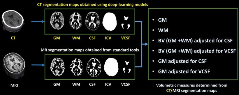
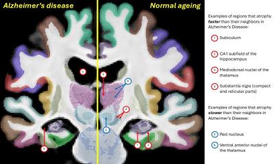
Image source: Srikrishna et al., Alzheimer's & Dementia 2023 (CC BY-NC-ND 4.0)
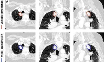
The new software can provide diagnostic support for radiologists and other professionals who interpret CT images. It has been created using deep learning, a form of artificial intelligence (AI). The software has been trained to transfer interpretations from MRI images to CT images of the same brains.

Image source: University of Gothenburg
“Our method generates diagnostically useful data from routine CT scans that, in some cases, is as good as an MRI scan performed in specialist healthcare,” says Michael Schöll, a professor at Sahlgrenska Academy who led the work involved in the study, carried out in collaboration with researchers at Karolinska Institutet, the National University of Singapore, and Lund University. “The point is that this simple, quick method can provide much more information from examinations that are already carried out on a routine basis within primary care, but also in certain specialist healthcare investigations. In its initial stage, the method can support dementia diagnosis, however, it is also likely to have other applications within neuroradiology.”
This is a well-validated clinical application of AI-based algorithms, and has the potential to become a fast and reliable form of decision-making support that effectively reduces the number of false negatives. The researchers believe that this solution can improve diagnostics in primary care and thus optimize patient flow to specialist care.

Image source: University of Gothenburg
“This is a major step forward for imaging diagnosis,” says Meera Srikrishna, a postdoctor at the University of Gothenburg and lead author of the study. “It is now possible to measure the size of different structures or regions of the brain in a similar way to advanced analysis of MRI images. The software makes it possible to segment the brain’s constituent parts in the image and to measure its volume, even though the image quality is not as high with CT.”
The software has been trained on images of a total of 1,117 people, all of whom underwent both CT and MRI imaging. The current study mainly involved healthy older individuals and patients with various forms of dementia. Another application that the team is now investigating is for normal pressure hydrocephalus (NPH).
With NPH, the team has obtained new results indicating that the method can be used both during diagnosis and to monitor the effects of treatment. NPH is a condition that occurs particularly in older people, whereby fluid builds up in the cerebral ventricular system and results in neurological symptoms. About two percent of all people over the age of 65 are affected. Because diagnosis can be complicated and the condition risks being confused with other diseases, many cases are likely to be missed. “NPH is difficult to diagnose, and it can also be hard to safely evaluate the effect of shunt surgery to drain the fluid in the brain,” continues Michael. “We therefore believe that our method can make a big difference when caring for these patients.”
The software has been developed over the course of several years, and development is now continuing in cooperation with clinics in Sweden, the UK, and the US together with a company, which is a requirement for the innovation to be approved and transferred to healthcare.
Source: University of Gothenburg
17.10.2023