

Image source: Blondis, MRI glioma 28 yr old male, marked as public domain, details at Wikimedia Commons
News • MRI & machine learning
A look into the genome of brain tumors
Researchers at Osaka University have developed a computer method that uses magnetic resonance imaging (MRI) and machine learning to rapidly forecast genetic mutations in glioma tumors, which occur in the brain or spine.
The work may help glioma patients to receive more suitable treatment faster, giving better outcomes. The research was recently published in Scientific Reports.
Cancer treatment has undergone a revolution in recent years. Spurred by recognition that each cancer case is unique, the specific genetic mutations tumor cells carry are now sequenced to discover which chemotherapy drugs will work best. However, certain types of cancer, especially brain tumors, are less accessible for genetic testing. The tumor’s genotype can’t be found until a sample is taken during surgery, and this can significantly delay treatment.
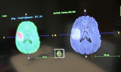
Glioma is a type of cancer that originates in the brain’s supporting cells. Two types of mutations are especially important; these are changes in the gene for the enzyme isocitrate dehydrogenase (IDH) or the promoter region of telomerase (TERT). Identifying these mutations can help direct the proper course of treatment. The researchers produced a machine-learning algorithm that can predict which mutations are present using only the MR images of the tumors. “Machine learning is increasingly used to diagnose medical images. But our work is one of the first to even attempt to classify something as hidden as the genotype based on image data alone,” study first author Ryohei Fukuma explains. The algorithm was found to be significantly better at predicting the mutations compared with conventionally used radiomic features of the MR images, such as size, shape, and intensity.
We hope to expand this approach to other types of cancer, so we can take advantage of the large cancer gene databases already collected
Haruhiko Kishima
To construct the algorithm, the researchers used a convolutional neural network to extract features from the MR images. Then, using a machine-learning method called support vector machines, they classified the patients into groups based on the presence or absence of mutations. “We hope to expand this approach to other types of cancer, so we can take advantage of the large cancer gene databases already collected” senior author Haruhiko Kishima says. The end result could remove the need for surgical tissue sampling. Even more, it could lead to better clinical outcomes for patients as the process of delivering personalized medicine becomes easier and faster.
Source: Osaka University
10.01.2020

