Image source: UCL
News • Coronavirus imaging
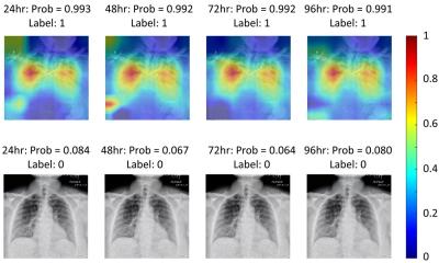
Brightest ever X-ray shows lung damage from Covid-19
The damage caused by Covid-19 to the lungs’ smallest blood vessels has been intricately captured using high-energy X-rays emitted by a special type of particle accelerator.
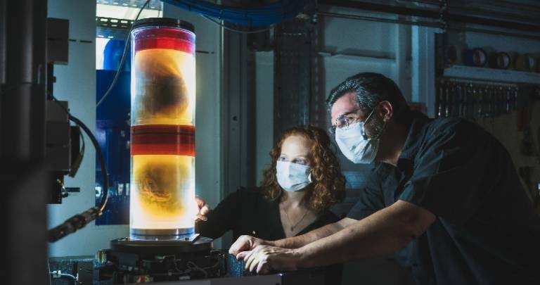
Scientists from University College London (UCL) and the European Synchrotron Research Facility (ESRF) used a new imaging technology called Hierarchical Phase-Contrast Tomography (HiP-CT), to scan donated human organs, including lungs from a Covid-19 donor.
The researchers published their finding in the American Journal of Respiratory and Critical Care Medicine; details on the new technology are available in the journal Nature Methods.
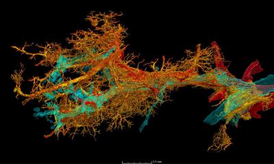
HiP-CT enables 3D mapping across a range of scales, allowing clinicians to view the whole organ as never before by imaging it as a whole and then zooming down to cellular level. The technique uses X-rays supplied by the European Synchrotron (a particle accelerator) in Grenoble, France, which following its recent Extremely Brilliant Source upgrade (ESRF-EBS), now provides the brightest source of X-rays in the world at 100 billion times brighter than a hospital X-ray. Due to this intense brilliance, researchers can view blood vessels five microns in diameter (a tenth of the diameter of a hair) in an intact human lung. A clinical CT scan only resolves blood vessels that are about 100 times larger, around 1mm in diameter.
By combining our molecular methods with the HiP-CT multiscale imaging in lungs affected by Covid-19 pneumonia, we gained a new understanding how shunting between blood vessels in a lung’s two vascular systems occurs in Covid-19 injured lungs
Danny Jonigk
Dr Claire Walsh (UCL Mechanical Engineering) said: “The ability to see organs across scales like this will really be revolutionary for medical imaging. As we start to link our HiP-CT images to clinical images through AI techniques, we will - for the first time - be able to highly accurately validate ambiguous findings in clinical images. For understanding human anatomy this is also a very exciting technique, being able to see tiny organ structures in 3D in their correct spatial context is key to understanding how our bodies are structured and how they therefore function.”
Using HiP-CT, the research team, which includes clinicians in Germany and France, have seen how severe Covid-19 infection ‘shunts’ blood between the two separate systems – the capillaries which oxygenate the blood and those which feed the lung tissue itself. Such cross-linking stops the patient’s blood from being properly oxygenated, which was previously hypothesised but not proven. Maximilian Ackermann MD (University Medical Center Mainz), clinical user of the technique, said: “Shortly after the beginning of the global pandemic we demonstrated that Covid-19 is a systemic vascular disease using histopathological (optical imaging of tissue) and molecular methods. However, these techniques did not adequately address the extent of the changes and clotting in fine blood vessels of whole lungs.”
Danny Jonigk, Professor of Thoracic Pathology, (Hannover Medical School, Germany) said “By combining our molecular methods with the HiP-CT multiscale imaging in lungs affected by Covid-19 pneumonia, we gained a new understanding how shunting between blood vessels in a lung’s two vascular systems occurs in Covid-19 injured lungs, and the impact it has on oxygen levels in our circulatory system."
Dr Paul Tafforeau, lead scientist at ESRF, said: “The idea to develop this new HiP-CT technique came after the beginning of the global pandemic, by combining several techniques that were used at the ESRF to image large fossils, and using the increased sensitivity of the new Extremely Brilliant Source at the ESRF, ESRF-EBS. This allows us to see in 3D the incredibly small vessels within a complete human organ, enabling us to distinguish in 3D a blood vessel from the surrounding tissue, and even to observe some specific cells. This is a real breakthrough, as human organs have low contrast and so are very difficult to image in detail with the current available techniques. ESRF-EBS has allowed us to go from deciphering the secrets of fossils to seeing the human body as never before."
Using HiP-CT to create the Human Organ Atlas
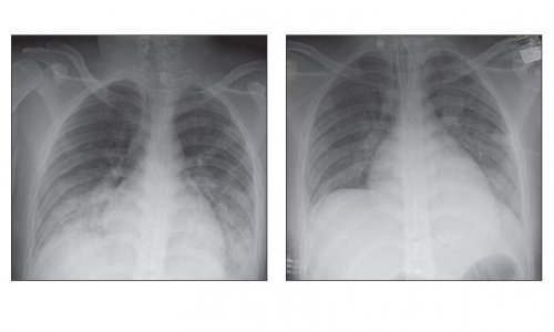
With support from the Chan Zuckerberg Initiative (CZI), the UCL-led team are using HiP-CT to produce a Human Organ Atlas, launching today. This will display six donated control organs: brain, lung, heart, two kidneys and a spleen, and the lung of a patient who died of Covid-19. There will also be a control lung biopsy and a Covid-19 lung biopsy. The Atlas will be available online for surgeons, clinicians and the interested public.
Project lead Professor Peter Lee (UCL Mechanical Engineering) said: “The Atlas spans a previously poorly explored scale in our understanding of human anatomy, which is the centimetre to micron scale in intact organs. Clinical CT and MRI scans can resolve down to just below a millimetre, whilst histology (studying cells / biopsy slices under a microscope), electron microscopy (which uses an electron beam to generate images) and other similar techniques resolve structures with sub-micron accuracy, but only on small biopsies of tissue from an organ. HiP-CT bridges these scales in 3D, imaging whole organs to provide new insights into our biological makeup.”
The researchers are confident that the scale-bridging imaging from whole organ down to cellular level could provide additional insights into many diseases such as cancer or Alzheimer’s Disease. Clinician Willi Wagner, Radiologist at University Hospital in Heidelberg said: “HiP-CT is filling a vast imaging gap in human medicine: clinical imaging provides 3D data of the body and organs but is limited to a gross scale; histopathology on the other hand provides detailed images of tissues and cells derived from small pieces of organs. It is generally limited to a small field and two dimensions. HiP-CT is bridging the organ to tissue scale, tightly linking the clinical disciplines of radiology and pathology and providing never before seen structural data of 3D tissue architecture and disease patterns.”
The authors hope the Human Organ Atlas will eventually contain a library of diseases that affect organs on a range of scales, from 1 to 100s of microns to entire organs, helping clinicians as they diagnose and treat a wide range of diseases. The team also hope to use machine learning and artificial intelligence to calibrate clinical CT and MRI scans, enhancing the understanding of clinical imaging and enabling faster and more accurate diagnosis.
Source: University College London
05.11.2021