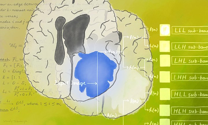
© Mindy Takamiya/Kyoto University iCeMS (CC BY 4.0)
News • Glioma grading
AI enhances brain tumour diagnosis
A highly accurate machine learning tool could help doctors tailor individualized treatments for people with glioma brain tumours.
A new machine learning approach classifies a common type of brain tumour into low or high grades with almost 98% accuracy, researchers report in the journal IEEE Access. Scientists in India and Japan, including from Kyoto University’s Institute for Integrated Cell-Material Sciences (iCeMS), developed the method to help clinicians choose the most effective treatment strategy for individual patients.
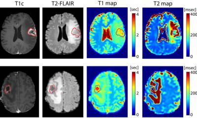
Gliomas are a common type of brain tumour affecting glial cells, which provide support and insulation for neurons. Patient treatment varies depending on the tumour’s aggressiveness, so it’s important to get the diagnosis right for each individual. Radiologists obtain a very large amount of data from MRI scans to reconstruct a 3D image of the scanned tissue. Much of the data available in MRI scans cannot be detected by the naked eye, such as details related to the tumour shape, texture, or the image’s intensity. Artificial intelligence (AI) algorithms help extract this data. Medical oncologists have been using this approach, called radiomics, to improve patient diagnoses, but accuracy still needs to be enhanced.
Recommended article

Article • Radiology + data + AI = ?
Today and future radiomics
Radiomics is one of the most exciting topics in radiology. It involves data and artificial intelligence (AI) but very few people know or understand the details. In her lecture ‘How does Radiomics work?’, presented at the German Radiology Congress in Leipzig, Professor Ulrike Attenberger outlined how radiomics will advance radiology but also the obstacles faced along the way.
iCeMS bioengineer Ganesh Pandian Namasivayam collaborated with Indian data scientist Balasubramanian Raman from Roorkee to develop a machine learning approach that can classify gliomas into low or high grade with 97.54% accuracy. Low grade gliomas include grade I pilocytic astrocytoma and grade II low-grade glioma. These are the less aggressive and less malignant of the glioma tumours. High grade gliomas include grade III malignant glioma and grade IV glioblastoma multiforme, which are much more aggressive and more malignant with a relatively short post-diagnosis survival time. The choice of patient treatment largely depends on being able to determine the glioma’s grading.
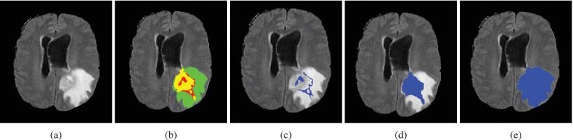
Image source: Kumar et al., IEEE Access 2020 (CC BY 4.0)
The team, including Rahul Kumar, Ankur Gupta and Harkirat Singh Arora, used a dataset from MRI scans belonging to 210 people with high grade gliomas and another 75 with low grade gliomas. They developed an approach called CGHF, which stands for: computational decision support system for glioma classification using hybrid radiomics and stationary wavelet-based features. They chose specific algorithms for extracting features from some of the MRI scans and then trained another predictive algorithm to process this data and classify the gliomas. They then tested their model on the rest of the MRI scans to assess its accuracy. “Our method outperformed other state-of-the-art approaches for predicting glioma grades from brain MRI scans,” says Balasubramanian. “This is quite considerable.” “We hope AI helps develop a semi-automatic or automatic machine predictive software model that can help doctors, radiologists, and other medical practitioners tailor the best approaches for their individual patients,” adds Ganesh.
Source: Kyoto University
05.06.2020