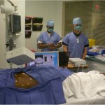
News • Treatment-resistant tumours
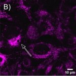
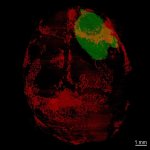
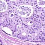
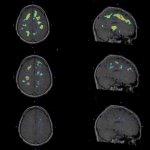
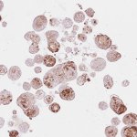
Researchers tear off "invisibility cloak" of colon cancer
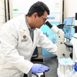
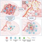
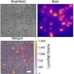
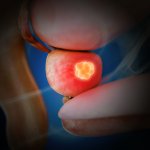
A study from the University of Calgary shows that removing a single gene makes colon cancer cells a target for immunotherapy — a fundamental breakthrough.


A study from the University of Calgary shows that removing a single gene makes colon cancer cells a target for immunotherapy — a fundamental breakthrough.
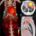
Radiopharmaceutical therapy has already transformed care for neuroendocrine tumors and prostate cancer, but other tumor types still lack targeted treatment options. A new approach could change this.
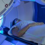
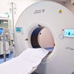
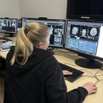
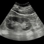
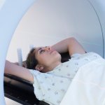
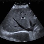
Imaging pregnant women with acute abdominal pain remains a significant clinical challenge – not only because of concerns about the impact on the unborn child, but also due to the anatomical changes that can obscure diagnosis. At the European Congress of Radiology (ECR) in Vienna, radiologists Vikas Shah and Charis Bourgioti examined the protocols for safe imaging in pregnancy and the management…
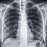
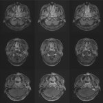
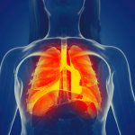
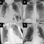
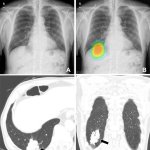
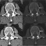
Researchers developed a simple, rapid, and low-dose X-ray technique to evaluate the severity of pulmonary valve regurgitation – a common complication after surgical repair of Tetralogy of Fallot.

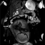
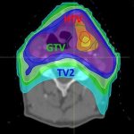
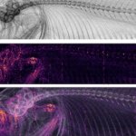
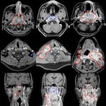
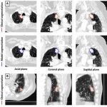
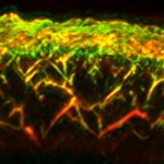
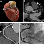
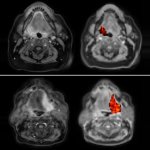
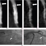
Researchers have produced the first 3D map of the heart’s electrical wiring in Tetralogy of Fallot, revealing features that may explain why many patients develop heart conduction disorders.
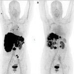
A new type of peptide receptor radionuclide therapy (PRRT) appears to be safe in metastatic neuroendocrine tumor patients who have exhausted conventional treatment options.

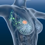
Obesity may change how early-stage breast cancer becomes invasive, according to a new study. The findings could help improve physicians’ ability to predict and treat the disease.

Due to advances in radiation therapy, some breast cancer patients may be able to omit surgery following ablative radiation, according to new results of a Phase 2 trial.

Once breast cancer begins to metastasise, it generally becomes harder to treat and survival rates are lower. Now, researchers present a new, targeted treatment approach using SBRT radiotherapy.
United Imaging debuts its CE-marked uRT-linac 506c at ESTRO 2026 – a CT-linac system with native AI architecture for adaptive radiotherapy workflows.
Artificial intelligence is enabling radiologists to extract valuable diagnostic information from routine chest imaging – identifying patients at risk for osteoporosis and cardiovascular disease without additional scans. At RSNA 2025, researchers presented two AI-powered tools that transform standard chest X-rays and CT scans into opportunistic screening devices.
Researchers are developing nanozymes to improve treatment of aggressive brain tumours. The tiny particles can be activated by near-infrared light and applied directly during surgery.
A new analytical method could improve how cancer treatments are designed – by allowing scientists to track, for the first time, exactly where inside a living cell a drug accumulates.

RFID – the same wireless technology that can track pets or locate items – can also be used to measure breathing in patients with impaired lung function contactlessly – in hospital or at home.

For a young adult, a cancer diagnosis hits different: a more aggressive disease course, greater disruptive potential, longer survivorship. Yet most healthcare institutions seem poorly prepared for this growing patient group. A plenary session at the NCCN 2026 Annual Conference examined a striking shift in modern oncology: the rising incidence of cancer in adolescents and young adults (AYA).
AI models can generate more complete summaries of complex cancer pathology reports than physicians, according to a new study that tested six models developed by Meta, Google, DeepSeek and Mistral AI.

The chances of breast cancer recurring remain very low when patients are treated with radiotherapy that is tailored to their individual risk following chemotherapy and surgery, new study results find.

Detector technology gets most of the attention in modern CT systems – but a new whitepaper by Dunlee, presented at ECR 2026, argues that the X-ray tube is equally decisive.
Two new studies explore the role of the thymus – a small organ in the chest, and possibly a missing piece in explaining why people age differently, and why cancer treatments fail in some patients.

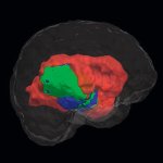
Researchers discovered that pineoblastoma, retinoblastoma and medulloblastoma – severe brain tumours in children that appear to be completely different – actually arise from the same type of cell.

The concept of using radiotherapy for osteoarthritis may seem counterintuitive for many clinicians. Yet a well-designed randomised controlled trial presented at the 2025 ASTRO Annual Meeting in San Francisco suggests that low-dose radiotherapy deserves a closer look. The findings add robust evidence to a therapeutic approach that has long been underutilised outside of German-speaking countries.

Radiation oncology is a field in remarkable transformation: a deepening global shortage of trained practitioners, persistent inequities in access to treatment – and, on the other side of the ledger, a new generation of technologies, from AI-driven adaptive planning to photon-counting CT, that are expanding what the field can do in ways previously unimaginable. At this year’s World Health Expo…

Functional brain radiosurgery is an application of stereotactic radiosurgery (SRS), representing its newest clinical field. It is a precise, non-invasive medical technique using focused ionising radiation to precisely target specific brain structures to modulate brain function for neurological disorders, psychiatric conditions, or intractable pain. The technology offers “precision without…
Omni Legend is a PET/CT total body scanning with 128 cm configuration. The remarkable scalable digital detector design at its core enables an unparalleled increase in true NEMA sensitivity without Lutetium intrinsic background radiation. Beyond its exceptional digital detector design, Omni 128 cm also delivers vast improvements to the entire PET/CT scanning process, such as more comfortable…
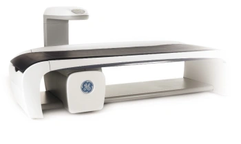
The DXA premium system covers the full range of clinical applications and provides detailed body composition analysis, including the distribution of fat, muscle, bone, and visceral fat. Its direct-digital HD detector delivers distortion-free visualization of bone structures without magnification errors. SmartScan reduces radiation dose and examination time, while Composer automatically generates…
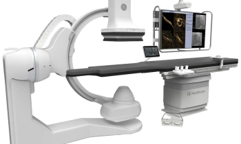
Allia IGS 5 Pulse is an interventional angiography system for radiology, cardiology, and neurology, featuring a newly developed user interface as well as a more powerful and quieter X-ray tube. Thanks to AI-based technologies for advanced image quality, such as CleaRecon DL, and the DoseCockpit, which continuously measures and adapts image quality and dose in real time, the system delivers…
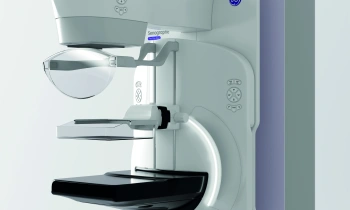
The Pristina Via from GE HealthCare is an innovative mammography system designed to enhance patient comfort and support efficient workflows for clinicians. Its modern, ergonomic design, intuitive interface, and flexible positioning enable a smooth and stress-free examination experience. The system can be optionally equipped with Dueta, allowing patients to actively participate in controlling…

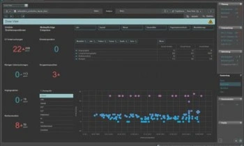
RadCentre Analytics offers an integrated solution for specific data analysis and interactive reporting to increase performance in radiology.Predefined and high performant processing of operating figuresUnlimited analysis options for optimisation of business outcomesIntegrated data warehouse solutionVisualization of radiation exposure extracted from PACS
RadCentre Dose View is a stand-alone and RIS-independent dose management system to assess patient exposures due to ionizing radiation. The system is able to meet legal requirements by offering consistent standards to increase the quality of radiological examinations.

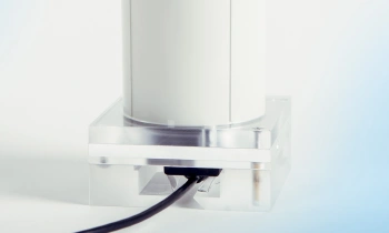
The Quart didoNEO introduces a new approach to diagnostic X-ray meters: it features the most compact base unit and most compact detector in the X-ray meter industry. The didoNEO R is used for QA and service in Radiography, (Pulsed) Fluoroscopy, DSA, Dental, 3D (CBCT).Compact multi-functional state-of-the-art solid state detectorEnables measurements in spots with limited spaceMeasures behind…
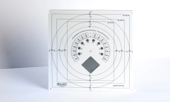
The Quart RFP150 phantom enables assessment of digital X-ray equipment according to the German DIN 6868-150 and DIN 6868-4.A small phantom version (the QUART SPdl) is available for fluoroscopy.The phantom can be ordered with a unique kV test object to routinely evaluate radiation quality and generator performance.Optional accessories include a suspension system for use on wall-mounted X-ray…
QUART dent/digitest 2D dental test phantoms are designed to assess X-ray imaging parameters according DIN and IEC QA / QC requirements.Features patient equivalent filtration and test objects to perform full-scale X-ray image quality analyses.Test Parameters are: Spatial resolution, High-contrast resolution, Low-contrast resolution, Homogeneity / artefacts, Radiation field / tube alignment
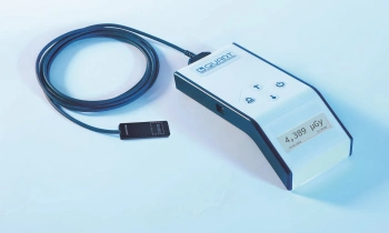
The Quart didoEASY meters are designed for quick measurements of dose, dose rate and exposure time in X-ray QA / QC and service.didoEASY meters automatically compensate all radiation qualities in their area of application. Three meter versions are available: for R/F and dental (50 – 150 kV), for mammography (25 – 40 kV), and one for the full diagnostic range (25 – 150 kV).
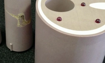
Our core competence is the development and production of customized phantoms in cooperation with our customers.We successfully collaborate with manufacturers in medical and industrial X-ray markets as well as with scientists and physicians working on research projects and studies.All standard phantoms can be modified according to your needs.We also offer customized phantoms for: PET, SPECT,…
VGi evo ensures a broad range of FOVs for acquisitions up to 24 × 19 cm. Volumetric, panoramic and teleradiographic exams as well as dynamic X-rays are available. Excellent image quality with very low radiated doses safeguards the patient’s health. A single scan generates HiRes images of airways, both TMJs, maxillary and nasal sinuses. Clear, precise scans reveal greater details of…
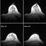
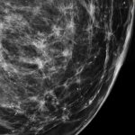
New research shows that using ultra-low field (ULF) MRI for breast imaging could offer an alternative to existing breast cancer screening methods and may reduce barriers to screening.

Radiotherapy is more effective when administered at the right time of day, according to new research. This discovery opens the door to cancer “chronotherapy”, the researchers hope.
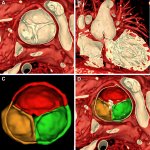
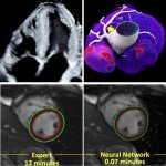
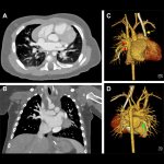
Researchers have developed 3D volume rendering methods for cardiac MRI that display complex structures within the heart and show blood flow. This can guide treatment decisions in pediatric patients.
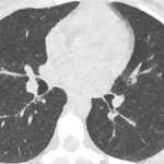
A study of 200 lung cancer patients shows photon-counting CT cuts radiation by 66% and contrast agent by 27% - while delivering sharper images and better tumor detection than conventional CT.
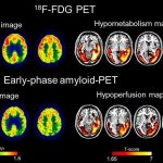
Beyond "one-size-fits-all": A new strategy that combines two types of PET scans can guide personalized radiotherapy for head and neck cancers, according to new research.

In today’s radiology environment, speed alone is not enough. What truly matters is intelligent workflow, diagnostic confidence and consistent performance even under pressure. Samsung’s GC85A Vision+ is designed exactly for that reality.

A new study shows a significant survival benefit for patients with oropharyngeal cancers treated with proton therapy (IMPT) compared to those treated with traditional radiation therapy (IMRT).

Researchers have discovered that low physical activity is associated with a higher risk of lymphedema. They have also noted that a lymph scanner objectively measures changes in the condition.

Radiotherapy is effective against prostate cancer but can cause side effects. Using AI, scientists found that images originally taken to help position patients could also predict rectal bleeding.
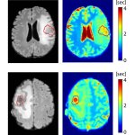
A novel AI-based method can distinguish between progressive brain tumours and radiotherapy-induced necrosis on advanced MRI. This could help clinicians more accurately identify and treat the issues.
At the RSNA 2025 annual meeting, Royal Philips has announced the expanded commercial availability of their LumiGuide 3D Device Guidance system for light-based navigation.

IDH mutated gliomas are slow-growing brain tumors with a relatively good prognosis. A new study shows that many patients reveal measurable cognitive impairment in the first year after treatment.
At this year’s RSNA annual meeting, Siemens Healthineers is presenting its new AI-powered solution for imaging procedures, called Optiq AI, designed to deliver higher quality low-dose images.
While saving lives remains the primary goal, a new study explores how radiation therapy also helps breast cancer survivors return to work. Policymakers should factor this in, the researchers argue.
Researchers developed an X-ray imaging method capable of revealing hidden features in a single shot. This could advance cancer detection, disease monitoring, security screening and material analysis.

Dunlee will present its portfolio of integrated imaging solutions at RSNA 2025 in Chicago, Illinois. The company will demonstrate technologies for diagnostic and therapeutic imaging applications, including developments in Ultra-High Resolution and Photon Counting CT (UHR & PCCT), components for MRI-guided breast biopsies, and onboard imaging systems for radiation therapy.

Researchers have developed a novel 3D-printable material that is both biocompatible and highly elastic. The technology could pave the way for artificial organs and improved drug delivery systems.

Researchers identified a targeted way to protect the brain from harmful side effects of cranial radiation therapy, potentially preserving the quality of life for millions of brain cancer survivors.
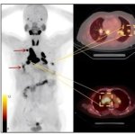
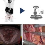
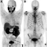
A novel imaging solution, called PSMA PET scanning, can more effectively detect the recurrence of prostate cancer compared to standard methods, and is associated with improved survival outcomes.
Cardiac imaging is evolving, and new techniques continue to uncover the secrets of the heart for cardiologists who know how to use them. At the ESC 2025 Congress in Madrid, four experts explored cutting-edge developments across different modalities. Ranging from AI-assisted ultrasound image acquisition and accelerated MRI protocols to advanced prognostic tools for CT and nuclear imaging, these…

At the EANS neurosurgery congress in Vienna, Italian medical imaging company Esaote presented I-Genius, a new open MRI system designed to provide real time checks during glioma surgery.
Scientists have shown for the first time that glioblastoma—the deadliest form of brain cancer—affects not just the brain but also erodes the skull, alters the makeup of skull marrow, and interferes with the body’s immune response.
Study of 3.7 million children reveals small but significant increased risk of blood cancers from medical imaging radiation, with CT scans posing highest risk

Hospitalists face a dual challenge when a critically ill pregnant patient is admitted to a hospital: providing safe and effective treatment for both mother and fetus. At the annual meeting of the Society of Hospital Medicine (SHM) in Las Vegas, an expert discussed why hospitalists must draw on a variety of skills when treating pregnant inpatients.

A newly developed method to accurately quantify how much radiation is absorbed by the blood during cancer treatment could lead to more personalised, preventive, and safer radiotherapy.
Patient communication facilitated by chatbots, image quality optimized by machine learning: Artificial intelligence (AI) is entering radiology at breakneck speed, transforming the specialty almost beyond recognition. So, how will the future of diagnostic imaging under AI look like, and which role will humans still play in it? At the ECR congress in Vienna, experts explored the societal and…
Tracer molecules serve to provide visual guidance in nuclear medicine, but a new molecule could give surgeons additional audio cues to help them locate prostate cancer tumors and metastases.
Gadolinium-based contrast agents enhance visibility but also pose a significant health risk. A new AI-powered virtual MRI imaging technique is designed to offer a safer diagnostic approach.

Classifying prostate cancer as “low-grade” could create a false sense of security and delay definitive treatments. A new study shows that the biopsy grade alone can paint an incomplete picture.
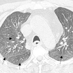
CT imaging is important to detect residual lung abnormalities after a Covid-19 infection. To avoid confusion with interstitial lung diseases, experts from 14 countries published a best-practice guide.
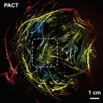
A new technique called photoacoustic computed tomography (PACT) offers a breast imaging alternative without the discomfort, high costs, or risk associated with the conventional evaluation methods.
A research group at Osaka Metropolitan University developed an AI model that can detect the presence of hepatic steatosis (fatty liver disease) from chest X-ray images.
Molecular profiling of over 1,000 nasopharyngeal cancer (NPC) tumours reveals distinct differences in tumour microenvironment of locoregionally advanced NPC, supporting personalised treatment.
Pulmonary embolism (PE) remains one of the leading causes of maternal mortality. At the French Thoracic Society Spring Days in May, Dr Aurélie Dehaene, radiologist at European Hospital in Marseille, France, reviewed diagnostic strategies for suspected PE during pregnancy, with a focus on clinical algorithms and optimized imaging protocols.
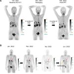
New research makes a strong case for a new dual-targeting radiopharmaceutical, designed to attach to two vulnerable sites on cancer cells, enabling more precise and potent therapy.
Ground-breaking innovation for endovascular surgery: The new software package Endovascular Navigation supports physicians in their interventions with planning, navigation and fusion imaging.

Lung cancer is the leading cause of cancer death in the EU, yet no organized screening program exists to detect the disease before symptoms appear. This September, France will strike back with an ambitious pilot program that could boost European lung cancer screening. Professor Marie-Pierre Revel presented the details at the French Thoracic Imaging Society Spring Days in Marseille, highlighting…

What if every radiographer could help combat climate change while performing their daily work? Following the congress theme of ECR 2025, experts revealed how small changes – from education initiatives to simple workflow adjustments – can collectively transform the environmental impact of radiology.

Nuclear medicine specialist Daniela Oprea-Lager has been appointed Professor of Theranostics at Radboudumc / Radboud University. Her research focuses on the combination of diagnostics and treatment using radioactive substances, with particular interest for urological tumors, especially prostate cancer.
With the introduction of boron neutron capture therapy (BNCT), Helsinki University Hospital offers a new radiation therapy method that can destroy cancer cells while sparing the surrounding tissue.

Integration of large language models (LLMs) in healthcare is still in its early stages, but expected to expand rapidly. Now, researchers point out potential cybersecurity vulnerabilities in radiology.
From organ-preserving treatments to chemo-immunotherapy combinations: five studies presented at ESTRO 2025 showcase how radiotherapy is reshaping the treatment landscape for anal and rectal cancer.

Bringing diagnostics and therapy closer together - that is the basic concept behind theranostics. Through further development of its PET/MR scanners, Siemens Healthineers aims to advance this approach. Andreas Schneck, Head of the MRI Division at Siemens Healthineers, talks about the new system, which was also presented at the European Congress of Radiology (ECR) in Vienna, and its advantages in…

The incidence of colorectal cancer in young adults has doubled over the last 20 years, with no known reason. Now, a new study links this mysterious trend to childhood exposure to a bacterial toxin.
Radiation therapy is an effective component of many cancer treatments, but some patients experience severe side effects. A new study shows that hyperbaric oxygen therapy can provide long-term relief.

Breast MRI has emerged as a powerful diagnostic tool, particularly for women with dense breast tissue where traditional mammography faces limitations. In her presentation at ECR 2025, radiographer Hanna Kalliomäki highlighted several technological advances transforming breast cancer detection and diagnosis. From time-saving abbreviated protocols and AI-assisted analysis to contrast-free…
Nuclear medicine (NM), one of the more mature technologies of diagnostic imaging, has been experiencing a rebirth in innovation and interest. The increasing prevalence of cancer,, an aging global population, and greater longevity, has created a robust demand for nuclear medicine. At ECR in Vienna, presenters explored market perspectives, but also safety and sustainability challenges.
Radiation safety experts developed a multi-purpose detector with a wide range of tools for different uses into a small package. For example, the detector detects all four kinds of ionizing radiation.

Surgery may not be the best next course of treatment for patients with early-stage breast cancer who had a complete response to neoadjuvant chemotherapy and standard radiotherapy, new research finds.
Agios Andreas General Hospital of Patras, Greece, is set to enhance its diagnostic capabilities with the installation of a new digital PET/CT imaging system.
To reduce the radiation exposure for patients undergoing frequent CT scans for pneumonia diagnosis, deep learning-based denoising of ultra-low dose CT presents a viable alternative.
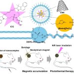
Japanese researchers have developed magnetic nanoparticles that can be directed to tumors using a magnet and then heated with a laser to destroy cancer cells while sparing healthy tissue.

United Imaging, a global leader in manufacturing advanced medical imaging and radiotherapy equipment, showcases a range of cutting-edge technologies aligned with sustainable values. Notably, the company achieves an A rating in the MSCI ESG ratings, highlighting its unwavering commitment to transforming medical diagnostics and patient care worldwide through a holistic, sustainable approach.

Photon counting detectors along with novel algorithms allow for more precise 3D visualization of different tissues and contrast agents by capturing X-rays at multiple energy levels simultaneously.

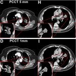
Pancreatic cystic lesions – indicating an increased risk of pancreatic cancer – are an occasional incidental finding in routine computed tomography (CT) abdominal imaging. New research suggests that the superior image quality of photon-counting CT (PCCT) can help detect more of these lesions. At the RSNA annual meeting, an expert outlined the benefits and limitations of the imaging technique…

For more than a year now, patients in Austria are no longer required to wear a lead apron during radiological imaging procedures such as X-ray and CT scans. However, the new recommendation, issued by the country's five specialist societies for radiation protection and imaging, has sparked mixed reactions among radiology technologists. At the annual congress of the specialist society…
The Diagnostica e Terapia Centro Aktis in Marano di Napoli, Italy, has expanded its diagnostic department with the addition of three imaging systems from United Imaging.

To improve breathing monitoring during radiotherapy, researchers have developed a millimeter-wave sensor capable of non-invasively visualizing respiratory movement during X-ray and CT examinations.
Researchers explore the connection between the gut and the brain and its impact on the onset of psychiatric and neurological disorders, including Alzheimer’s.
Late-stage breast cancer patients are more often affected by brain metastases than previously believed, a new study finds. The researchers suggest that MRI screening guidelines should reflect this.
Senescent cells, which may appear after chemotherapy or radiotherapy, can jeopardize patients' recovery. A study describes a new mechanism to eliminate these cells in cancer patients.
A new deep learning model shows promise in detecting and segmenting lung tumors. The findings of the study could have important implications for lung cancer treatment.
The installation of a state-of-the-art digital PET/CT scanner in Essen marks the German debut for the technology of United Imaging. The device is designed to deliver precision diagnostics at the Kliniken Essen-Mitte Evang and the private nuclear medicine clinic Nukmed, a premier cancer care institution.
Steam eliminates wrinkles and germs, but can it destroy cancer cells too? A multisite clinical trial explores the potential of a water vapor system using steam to kill prostate cancer cells.
Why does radiation therapy kill cancer cells from the same tumour in different ways? This has long remained poorly understood. Now, new findings open up opportunities to improve treatment.
A premiere that brings hope for fertility restoration to men who underwent chemotherapy during childhood: Researchers reintroduced cryopreserved immature testicular tissue taken 16 years prior.
United Imaging announces four installations of their diagnostic imaging systems at three healthcare facilities in Romania. Working closely with distributor Tehnoplus Medical, the company hopes to increase diagnostic accuracy and support better treatment outcomes.
Precise segmentation of anatomical structures greatly benefits cancer diagnosis. Using AI and deep learning methods, researchers are developing a high-precision 3D viewer software for medical image data.
A research team has found wearable organic x-ray sensors could be the answer towards safer radiotherapy protocols for cancer patients, reducing the debilitating side-effects of the treatment.
United Imaging are excited to share that, in partnership with Fora S.p.A., an industry leader with over 50 years of expertise in delivering advanced diagnostic technologies to the Italian market, Policlinico Casilino Hospital will join the company's global network. Demonstrating its commitment to innovation and growth, the hospital has selected the industry-leading 640-slice CT scanner to…
Royal Philips announced a major advance in radiation oncology with 510(k) clearance from the US Food and Drug Administration (FDA) for its new detector-based spectral CT radiotherapy solution.

Hyperbaric oxygen therapy (HBOT) treatments could offer relief to breast cancer patients who experience late toxicities following radiotherapy treatment. To date, the handful of completed clinical trials only produced inconclusive or contradictory results. Therefore, results from the latest trial, named HONEY (Effect of Hyperbaric Oxygen Therapy on Breast Cancer Patients with Late Radiation…

Recent developments in deep learning techniques are enhancing clinical imaging quality and reducing radiation exposure for patients while also maintaining diagnostic accuracy. The latest AI (artificial intelligence) component to clinical imaging – referred to as deep learning reconstruction (DLR) – is having a particular benefit in paediatric imaging, according to Dr Samuel Brady from…

New technology shows promise in protecting an implant against infections from resistant bacteria: By heating up small nanorods of gold with near-infrared light (NIR), the bacteria are killed.

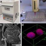
A ‘mini-protein’ can deliver radiation doses directly to tumours without harming healthy tissues. The approach shows promise for the treatment of metastatic bladder cancer and other tumours.
A new study found that measuring circulating tumor cells (CTCs), rare cancer cells shed from tumors into the blood, is a reliable way to predict later treatment response and survival prospects with metastatic prostate cancer.
A bit more mucus in the nose, a little less air in the gut: Even small changes can be important when planning proton therapy against cancer. A new workflow allows for an adapted irradiation every day.

Ultrasound plays a pivotal role in the assessment of organ transplant patients. It enables physicians to safely and easily assess progress, identify complications and resolve problems, as well as deliver long-term monitoring. The value of ultrasound in the transplant space was highlighted in a session at ECR 2024, covering liver, renal, pancreas, paediatric and multi-organ transplants with…
Researchers have identified fibrotic scarring as a key source of resurgence of glioblastoma multiforme (GBM). The new insights could lead to better prevention of this type of brain cancer.
Radiologists and gynaecologists entered a veritable bout during the Oxford debate at the 43rd annual congress of the German Senologic Society in Dresden. Controversy was sparked by the question of whether all patients with a ductal carcinoma in situ (DCIS) should undergo radiotherapy.
Radiotherapy for breast cancer is always associated with the risk of damage to organs or surrounding tissue. A new positioning system, which has now been patented by the Hamm-Lippstadt University of Applied Sciences (HSHL), positions the breast far away from the upper body and thus increases the distance to the organs at risk. The medical device called "X-Akt Mamma RTX" is now set to be…
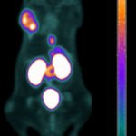
To diagnose neuroblastoma in children, lengthy scans and anaesthesia are often required. A new PET/CT imaging technique could deliver faster results without sedation for the paediatric patients.
United Imaging, a company specializing in advanced imaging solutions, is proud to announce the first installment of their cutting-edge technology in France. The increasing number of healthcare facilities deciding to leverage the company's diagnostic solutions stands as a testament to their effort toward engineering imaging solutions that hold the potential to transform patients’ lives worldwide.

Can a mouthwash-based test help predict head and neck cancer recurrence? A new study suggests it might.

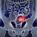
When a person is diagnosed with rectal cancer, part of the bowel is often removed, often resulting in the need for a stoma or problems with bowel control. A new therapy could help avoid this surgery.

New resesarch suggests a new way of assessing the risk of heart failure without invasive diagnostic tests. The method involves MRI to measure heart pressure as a predictive factor.
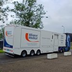
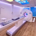
United Imaging is pleased to announce the installation of their uMI 550 mobile system in Germany. Starting in September, Alliance Medical Northern Europe (part of the Alliance Medical Group) is using the manufacturer’s advanced imaging technology to facilitate access to the highest quality diagnostic services for their patients. Germany is now the second country in Europe where the company's…
After breast cancer surgery, radiotherapy helps prevent the cancer from returning. However, this protection does not last forever. A long-term study shows the duration of the treatment's benefits.
Handheld point-of-care diagnostics, magnetic endoscopy, AI-enhanced robotic surgery, smart patient information management, wireless minimally invasive surgery systems, and much more: At the Medical Taiwan Health & Care Expo in Taipei this summer, visitors had the opportunity to see innovative medical products and solutions across a wide range of specialties. We took a closer look at selected…
As opportunities for teleoperations rapidly expand within radiology, the concept is being deployed across an array of modalities to deliver more efficient healthcare. A range of speakers covered the topic of ‘Teleoperations in radiology’ at ECR2024, discussing its benefits in applications in MRI, ultrasound, during the social restrictions of the Covid-19 pandemic and military use. However,…
Ductal Carcinoma in Situ (DCIS) often develop into more invasive forms of breast cancer. To predict which DCIS patients are likely to be affected, researchers have developed an analytic AI-based tool.

The nuclear medicine global market is projected to see a significant increase in the coming years, with the lion's share being attributed to radiotherapeutics. So, how to set up a dedicated theranostics centre? At the annual meeting of the Society of Nuclear Medicine and Molecular Imaging (SNMMI) in Toronto, Ontario, an entire session was dedicated to planning logistics, radiation safety, and…
When colorectal surgery was first performed with robotic assistance in 2014, the procedure was questioned about safety, efficacy, and outcomes. Today it is an established option. Well-trained surgeons use robotic surgical systems confidently. Numerous clinical studies have verified its intraoperative benefits for patients and surgeons alike, as well as very positive outcomes for patients.
Smaller kills faster – this is what was previously thought about gold nanoparticles used to fight cancer cells. However, new research reveals a more complex picture of these interactions.
A new radiotracer enables positron emission tomography (PET) scans to be used for the first time to accurately pinpoint when and where tuberculosis (TB) is still active in a patient’s lungs.

What is an advanced clinical practitioner (ACP) and why should nuclear medicine technologists strive to become one? At the 2024 annual meeting of the SNMMI in Toronto, ACP Luisa Roldão Pereira outlined the position and its importance in the clinical context.
Researchers demonstrated how the growth of malignant brain tumours can be greatly decreased by using iontronic technology to continuously administer low doses of cancer drugs.

The concept of delivering radiation therapy to cancer patients seated in an upright position is undergoing a major resurgence. Evidence is already highlighting that patients feel more comfortable seated upright and enjoy better communication with radiotherapists during their care. In addition, there are indications of less internal organ movement, enabling more accurate treatment delivery.…
The IRCCS in Bologna has inaugurated a state-of-the-art integrated PET/CT system. This cutting-edge technology allows for the entire human body to be studied in a single scan, even detecting the smallest tumour cells.
A combination of facial thermal imaging and artificial intelligence (AI) can accurately predict the presence of coronary artery disease (CAD), new research finds.
In more than one in six patients diagnosed with pancreatic cancer, the tumour is initially diagnosed at a non-metastatic primarily unresectable, locally advanced stage (LAPC). For these patients, a new internal radiation procedure, OncoSil™ brachytherapy, may become a treatment option – in Germany, around 858 patients could benefit from this innovation annually.
Diagnosing lung conditions such as pulmonary embolism is more challengenging when a patient cannot tolerate contrast agents. Now, a new software solution is addressing the issue.
Chemotherapy kills cancer cells – but how? New research suggests that the mechanisms are different than previously understood. The finding will have implications for future cancer treatments.
AI-enhanced CT scans can accurately evaluate cardiovascular risk without contrast media, a new study shows. The technique uses coronary calcium and heart chamber size as markers for disease detection.
In some patients with multiple sclerosis (MS), symptoms worsen during treatment, but MRI scans do not indicate any change. In such cases, positron emission tomography could help, a new study suggests.

Prostate radiotherapy techniques have been transformed over the past two decades. One promisting technique in this context is magnetic resonance-guided radiotherapy. The latest clinical results show a dramatic reduction in side effects, improving patient outcomes and quality of life.
A new trial could pave the way for more gentle surgery of breast cancer: The researchers explore the possibility of sparing the lymph nodes in the armpit - even if metastases are already present.

New approaches to cardiovascular radiology are evolving to help clinicians gain an increasingly better insight into heart conditions. Latest developments in cardiovascular radiology include myocardial strain imaging, 4D flow and photon-counting CT technology. An ECR 2024 session shone the spotlight on these areas of cardiovascular imaging with expert speakers outlining the pros and cons of each.

US oncologists are exploring a new combined chemotherapy and surgical approach to safely remove advanced pancreatic tumors that were previously considered inoperable.
A novel AI-based, non-invasive diagnostic tool enables accurate brain tumor diagnosis, outperforming current classification methods. The tool leverages MRI information to aid clinical decision making.

Holding a mobile phone close to the head for an extended amount of time has long been connected to brain cancer. Now, a new study found no hints for an increased risk.

Striking the balance between diagnostic efficacy and patient safety remains critical when utilising iodinated contrast media to deliver the best imaging outcomes. While playing a crucial role in diagnosis and treatment of disease, CT expert Efthimios Agadakos believes the medical profession has a duty to do its utmost to minimize patient risk from contrast media.
Dutch researchers use PSMA targeting to improve detection of prostate cancer, improving nodal staging and guiding more accurate surgery for this important patient population.
Stagnation, under-use, unfulfilled potential: At the EUSEM congress in Barcelona, leading emergency physician Dr Joseph Osterwalder describes how e-FAST (Extended Focused Assessment with Sonography for Trauma) – a key point-of-care ultrasound technique for trauma – has changed over the last two decades, and not necessarily for the better.

Cancer patients receiving radiotherapy run the risk of injuring their lungs. This can lead to conditions like pneumonitis and fibrosis. A new cell-by-cell model can help make treatments safer.
Using light instead of x-rays, a new imaging method from Philips is designed to advance navigation through a patient's blood vessels during minimally-ivasive procedures.

A newly-developed biosensor could be an alterative to MRI and mammography for breast cancer screening. The handheld device uses saliva on test strips to determine whether cancer is present.

On 9 December 2023, NovaLife Polyclinic, with over 15 years of experience in the private healthcare sector in Timișoara, proudly inaugurated its state-of-the-art branch in the vibrant capital city of Bucharest.
A new breast imaging technique provides high sensitivity for detecting cancer while significantly reducing the likelihood of false positive results, according to a new study.
Researchers in Singapore have developed an AI-based software to assist in the early detection of breast cancer. Using thermal imaging, the program assesses the malignancy of a tumour.

United Imaging Healthcare Europe, a leading company in advanced medical imaging and radiotherapy equipment, proudly announces the introduction of its first Mobile Digital PET/CT solution in Italy, now fully operational in the Piacenza province under the auspices of the Azienda Unità Sanitaria Locale Piacenza (AUSLP) hospital.
RTI presents its latest advancement in X-ray Quality Assurance and testing. Centered around efficiency, Mako provides the ultimate no-fuss experience, simple setup in all X-ray applications, and fully wireless capability.
Neuroscientists recently discovered that low-dose ionizing radiation (LDIR) can reduce lesion size and reverse motor deficits in TBI and ischemic stroke mice, demonstrating its therapeutic potential.

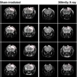
Using a unique new technique, US researchers hope to offer a safer and more effective alternative to current cancer treatments, reporting promising first results in mice.
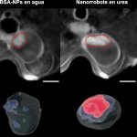
New research demonstrates how tiny nanomachines could greatly reduce bladder cancer by precisely targeting the tumour and attacking it with a radioisotope carried on their surface.
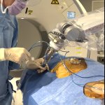
Long confined to surgery, robots are making their first steps in interventional radiology. Those devices could help improve accuracy in tumour targeting during needle insertion and help less experienced radiologists perform ablations, a leading French interventional radiologist showed at the Spectrum conference in Miami.
Using AI and optoacoustic imaging, researchers have developed a new method to assess microvascular changes in the skin – and thus the severity of diabetes in the patient.
University of Waterloo researchers are pioneering a method to detect breast cancer in women early enough for them to receive life-saving treatment.

New research suggests a new approach to precision radiotherapy can reduce the risk of swallowing problems for patients, without impacting the success of treatment.

Smart catheters, smart diapers or wound dressings: a new approach to wireless biosensors from Malmö University opens up options for more patient-controlled ways of infection detection.
Bone scans have been found to overstage prostate cancer at initial staging compared to prostate specific membrane antigen (PSMA) PET, according to new research.
Ultrasound technology now plays a vital role in clinical diagnosis and management. Significant advances in point-of-care ultrasound (POCUS) have made it a versatile tool for assessment, diagnosis, and follow-up across various fields. New developments continue to expand its applications, improving patient care and outcomes.
A multinational study confirms a strong and clear association between exposure to radiation from CT scans in young people and an increased risk of blood cancers.

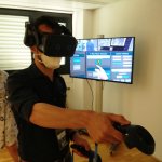
Giving children a virtual/augmented (mixed) reality playkit to use ahead of an MRI scan seems to ease both their and their parents’ anxieties about this procedure, a new study suggests.
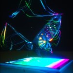
Inspired by the enhanced visual system of butterflies, researchers have developed an imaging sensor to “see” into the UV range for differentiating between cancer and normal cells.

Because time is brain: To explore the potential for accelerated stroke diagnostics, US researchers equipped an ambulance with a portable MRI – with promising first results.

Seeing a female doctor in a film may inspire women to pursue a career in medicine. However, the clinical gender reality is not well-represented in movies, a new study finds.
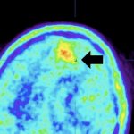
Advances in positron computed tomography (PET) could lead to a more refined approach to the precise removal of brain tumors is on the horizon, experts from Poland point out.
New trial results suggest that a short course of induction chemotherapy prior to chemoradiation could reduce the rate of relapse and death among patients with locally advanced cervical cancer.

A new research collaboration aims to develop a forward-looking AI application to detect gender-specific symptoms earlier and further reduce mortality from heart disease, especially among women.
High-BMI patients are a challenge for abdominal sonography. In a new study, researchers point out the benefits of special high-performance probes and their impact on image quality.
After pelvic radiotherapy, patients may live with low-grade chronic inflammation of the lower intestine 20 years after the treatment, a new study shows.
US researchers have discovered that radiation therapy combined with two types of immunotherapy can control tumors in preclinical models of triple negative breast cancer.
Reports of AI gaining the upper hand in diagnostic imaging interpretation are piling up, but there are still areas where the eye of a trained human radiologist remains superior.

Experts from Brigham and Women’s Hospital have tasked ChatGPT to generate recommendations for cancer treatment – with some promise, but ultimately inadequate results.

Experts presented state-of-the-art and emerging techniques to treat chest tumours and discussed common issues in the management of pneumothorax at RSNA 2022. Current ablation methods in the thorax include radiofrequency ablation (RFA), microwave ablation (MWA), cryoablation (CRYO), irreversible electroporation (IRE) and pulsed electric field.
In a new editorial paper, researchers from the Army Hospital Hamburg discuss treatment strategies for common, but highly malignant types of head and neck tumors.
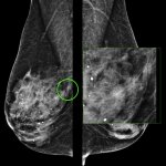
With the recent recommendation changes from the European Council in 2022, how radiologists screen for breast cancer is changing. Mammography has long been an essential technology in screening for breast cancer, and in the recommendations the Council formally recognized the advantages of digital breast tomosynthesis (DBT). This landmark acknowledges the research on and benefits of DBT, advocating…
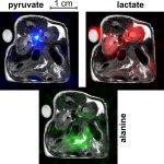
A new imaging method devised by German researchers offers a fast and cost-effective way to observe abnormal metabolic processes live in the MRI scanner.
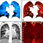
Photon-counting CT allows for a comprehensive, simultaneous evaluation of lung structure and function, something not possible with standard CT, according to a new study.

Should radiation dose from previous medical imaging be considered when a new scan is made? UK experts issued a joint position statement that favors clinical need over prior exposures.

Treatment times for radiotherapy could be reduced for some early breast cancer patients, according to a trial led by University of Cambridge and The Institute of Cancer Research, London.
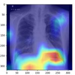
Assistance from an AI algorithm with high diagnostic accuracy improved radiologist performance in detecting lung cancers on chest X-rays and increased human acceptance of AI suggestions.

For proton radiation therapy against cancer, there is yet no direct method for mapping the beam range during dose delivery. A new method devised by Dresden scientists could help.
Photon-counting CT enables accurate diagnosis of coronary artery disease in high-risk patients, a potentially significant benefit for people previously ineligible for noninvasive screening.
US researchers evaluated AI techniques for cleaning medical images based on performance in clinical tasks. Some algorithms make the scans look better, but actually decrease image quality.

US researchers identified a potential breakthrough in glioblastoma treatment. Using a modified virus, they created a treatment that specifically attacks tumor cells, while leaving normal cells intact.

Against a backdrop of changing technology and reduced patient dose, a new momentum is emerging within radiology to eradicate patient shielding. The subject has been extensively debated and researched in recent years but there is now a growing consensus to end the practice, apart from with a few exceptions. The topic was the focus of a session at ECR 2023 in Vienna where different perspectives…
A new advanced form of CT imaging offers better cardiovascular imaging quality compared to dual-source CT (DSCT) in infants with suspected cardiac heart defects, according to a new study.
Researchers from the Organoid group (Hubrecht Institute) and UMC Utrecht have developed a biobank with organoids derived from patients with head and neck cancer (HNC).

Patients with early-stage breast cancer who have an elevated risk of having tumour recurrence now have the option to take a course of radiotherapy following breast conservation surgery that is only three weeks long, half the time of conventional radiotherapy treatment.
Stereotactic body radiotherapy (SBRT) is increasingly used in early-stage lung cancer. However, experts caution that ditching surgical options in favour of "SBRT-only" has serious drawbacks.

Artificial Intelligence (AI) could identify patients at increased risk of side effects from radiation treatment for breast cancer, according to researchers at the University of Leicester.

A new online ‘European Cancer Pulse’ tool, created to compare cancer data across Europe, has highlighted that only 12 of the 27 EU Member States have an up-to-date national cancer control plan.

For patients with human papilloma virus associated oropharynx cancer, assessing the presence of cancer cells beyond the lymph nodes is critical in determining proper treatment.
When is a CT scan justified, i.e. when do the benefits of a CT scan for the patient outweigh possible risks associated with radiation? Justification has been a major issue among radiologists ever since CT has become widely available and widely used. With regard to dose the answer is the well-known ALARA principle: “As low as reasonable achievable“. Now, the European coordinated action on…
For decades, researchers have marveled at the ability of glioblastoma, a particularly aggressive brain cancer, to turn off a patient's cancer-fighting immune cells, thereby allowing tumors to grow freely.

Women working in health care who are regularly exposed to radiation from X-rays and other imaging procedures need better ionizing radiation protection.

Dunlee unveiled its new oncology bundles onsite at ECR. The bundles combine components that have been tested and verified to work together so clinicians can offer state-of-the-art onboard Cone Beam CT (CBCT) in facilities.

A new clinical trial has shown that men diagnosed with prostate cancer can benefit from ‘radical radiotherapy’ that delivers treatment in five hospital visits instead of the typical 20.
A novel imaging modality that can visualize the distribution of medical radiopharmaceuticals with very fine resolution has been developed and successfully tested.

Chatbots become popular resources for cancer information - but are their results accurate? Researchers evaluated the reliability of ChatGPT’s cancer information.
Imaging researchers have taken a major step towards their ultimate goal of identifying cancers that are starved of oxygen so that altered treatment can be used to target them more effectively.

Radiology practitioners have highlighted the benefits of cone beam CT in delivering high resolution at a low dose. Delegates at ECR 2023 in Vienna heard how cone beam CT (CBCT) could replace multidetector CT (MDCT) in some areas and is already showing cost-effectiveness benefits.
Researchers at the University of Barcelona have developed a new tool to assess the presence and severity of sarcopenia, using an ultrasound-based muscle quality scoring system.

ECR 2023 returns to its traditional date in March, but delegates can expect novelties with sessions touching not just cutting-edge science, but also archaeology and palaeontology, and putting trainees in the spotlight, Congress President Professor Adrian Brady told Healthcare in Europe in an exclusive interview.

Radiation exposure in diagnostic and interventional radiology is steadily being reduced, but some important parameters have hardly been taken into account so far, says Dr Kerstin Jungnickel. The medical physics expert explains how patient-specific protocols can improve radiation protection and outlined new findings on the radiosensitivity of certain body regions and their impact.
Radiotherapy does not improve survival rates in older patients with early breast cancer, new research suggests.

The newly founded UK Focused Ultrasound Foundation is dedicated to advancing the development and adoption of the technology, which can be used to non-invasively treat tissue deep in the body.
A novel nuclear medicine imaging protocol can take the place of two separate imaging scans for the evaluation of brain changes linked to cognitive impairment.
Evidence that radioembolization, a trans-arterial therapy, is safe and stops disease progression in metastatic breast cancer is increasing, a prominent American interventional radiologist showed at the Spectrum conference in Miami.
A new study from Spain has demonstrated the efficiency of an ultrasound radiation-based therapy on the inhibition of cancer cell motion in pancreas cancer models.
Photon-counting detector CT reduces the amount of contrast needed for CT angiography (CTA) while maintaining image quality, according to a new study published in Radiology: Cardiothoracic Imaging.

Monitoring the proper blood supply to the brain could be used to prevent or even treat neurological diseases. A new technique called πNIRS aims to do just that.
In men recently diagnosed with intermediate or high-grade prostate cancer, PSMA PET/MRI can successfully determine whether their cancer is likely to return within two years of a prostatectomy.

Engineers have developed an electronic patch that can give medical professionals unprecedented access to crucial information that could help spot life-threatening conditions.

AI-based models for multimodality hybrid imaging have the potential to be a potent clinical tool but are currently held back by a lack of transparency and maturity, says Dr Irène Buvat, from the Laboratory of translational Imaging in Oncology, Institute Curie in Paris, France.
A promising new application for photon-counting CT: The new technology outperforms conventional CT in detecting subtle damage in the lungs of patients with persistent symptoms of Covid-19.
Canadian researchers have developed a new method of killing brain cancer cells while preserving the tissue around it. A remarkable side-benefit: chemotherapy of the cancer suddenly becomes possible.
Using fluorodeoxyglucose positron emission tomography (FDG-PET) imaging may give insights into possible dose reductions in ongoing radiation therapy of head and neck cancer. A promising study to explore this option was presented at the 2022 ASTRO/ASCO Multidisciplinary Head and Cancer Symposium held in Phoenix, Arizona.

Radiographers could help design new artificial intelligence (AI) tools for radiation protection, Mark McEntee, professor of diagnostic radiography at University College Cork, Ireland, argued during the annual EuSoMII meeting in October.

An advanced radiotherapy technique can be used to safely treat prostate cancer patients in as little as one to two weeks, compared with the current standard, which takes one to two months.
A new randomized study confirms that men with high-risk prostate cancer can be treated with a moderately shortened course (5 vs. 8 weeks) of radiation therapy.

Speed or accuracy? As far as Covid-19 tests go, this was the choice you had to make. In the future, this dilemma could be a thing of the past.
Bowel cancer patients could in future benefit from a new 3D bioprinting technology which would use their own cells to replicate the complex cellular environment of solid tumours in 3D models.
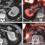
99mTc-Sestamibi SPECT/CT can aid in the diagnosis of solid renal lesions, especially in cases where contrast enhanced CT findings are inconclusive, according to researchers from the Mallinckrodt Institute of Radiology at Washington University School of Medicine in St. Louis, Missouri.

Focal treatment of prostate cancer involves treating only the tumor and not the entire gland, sparing surrounding tissue and nerves. The method is being evaluated in a research study.

Deaths from cancer are currently estimated at 10 million each year worldwide. Conventional cancer staging systems aim to categorize patients into different groups with distinct outcomes. ‘However, even within a specific stage, there is often substantial variation in patient outcomes,’ Markus Plass, academic researcher from the Medical University of Graz, Austria, explained to Healthcare in…
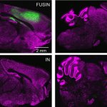
Treatment of central nervous system diseases and tumors is often hindered by the blood-brain barrier. A new method aims to overcome this obstacle using focused ultrasound intranasal delivery (FUSIN).

At this year's Medica (Nov 14-17), the Health IT Forum offers a special focus on how health IT can contribute to more sustainability in healthcare, and on optimising treatment workflows.
In the future, many types of open surgeries will be replaced with minimally invasive interventions, predicts Kevin Cleary, PhD, engineering lead at the Sheikh Zayed Institute for Pediatric Surgical Innovation at Children’s National Hospital, and Professor of Pediatrics and Radiology at George Washington University, both in Washington, D.C. Surgeons and interventional radiologists will be able…
US researchers have developed a test to detect loss of myelin - a key contributor to many neurological diseases including multiple sclerosis, traumatic brain and spinal cord injuries, stroke, and dementia.
Recent advances in CT have focused mainly on software, yet new technology could push the modality much further, experts showed at ECR 2022.
Researchers at Queen’s University Belfast have developed a new plastic film that can kill viruses that land on its surface with room light.

Ethical considerations continue to fuel the discussion around artificial intelligence (AI), a panel of experts showed at ECR 2022.

Machine learning and artificial intelligence (AI) have the potential to transform cancer treatment management worldwide. Their ability to rapidly analyse and integrate routinely acquired diverse data will improve the accuracy and effectiveness of precision medical treatments.
New CT technology paired with artificial intelligence-based noise reduction offers superior detection of bone disease associated with multiple myeloma at lower radiation doses than conventional CT.
UK researchers developed and validated a deep learning algorithm that can identify and outline a non-small cell lung cancer (NSCLC) tumor on a CT scan within seconds.
A surprising mechanism explains why high-grade gliomas, the deadliest form of brain cancer, returns: The tumours adapt to treatment by recruiting help from nearby healthy tissue.
Exposure to ionising radiation is a major health concern for many interventional radiologists. To help attain an understanding for radiation distribution in a clinical setting, experts tap into the potential of virtual reality (VR).
The combination of deep-learning reconstruction and a subtraction technique yields promising diagnostic performance for the detection of in-stent restenosis by coronary CTA.
A spin-off from ETH Zürich has developed a magnetically steerable catheter for quick and safe stroke treatment that no longer requires surgeons to be on site.

Researchers are seeking alternatives to gadolinium-based contrast agents (GBCAs) to help raise levels of patient safety. An open hybrid session at ECR in Vienna heard how research across several centres has been examining the options of new approaches to reduce reliance on GBCAs.

A Europe-wide project aims to improve medical radiation protection for patients and staff by better understanding and evaluating the health effects of exposure to low-dose ionising radiation resulting from diagnostic and therapeutic procedures.
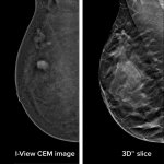
Contrast-enhanced mammography (CEM) has gone a long way since it was first approved by the Food and Drug Administration (FDA) in 2011. An Italian expert explained how useful the modality has become in breast imaging in a dedicated course at ECR 2022.