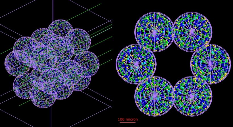
Image source: Cogno N et al., Communications Medicine 2024 (CC BY 4.0)
News • Research on radiation interaction
Lung tissue model to increase cancer radiotherapy safety
An innovative computer model of a human lung is helping scientists simulate, for the first time, how a burst of radiation interacts with the organ on a cell-by-cell level.
This research, carried out at the University of Surrey and GSI Helmholtzzentrum für Schwerionenforschung, Darmstadt, and published in the journal Communications Medicine, could lead to more targeted treatments for cancer and reduce the damage caused by radiotherapy. "Doctors could one day use our model to choose the right length and strength of radiotherapy – tailored to their patient. This is exciting enough – but others could use our technique to study other organs. This could unlock all kinds of medical knowledge and could be great news for doctors and future patients," says Dr Roman Bauer, Senior Lecturer at the University of Surrey.
[This model] will allow us to study the way fibrosis and other conditions are actually caused, and how they develop over time
Marco Durante
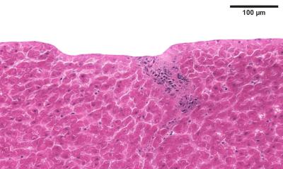
Nowadays, more than half of cancer patients receive radiotherapy – but too high a dose can injure their lungs. This can lead to conditions like pneumonitis and fibrosis. To study these injuries, researchers at GSI and the University of Surrey used artificial intelligence to develop a new model of part of a human lung – cell by cell.
"For the first time, BioDynaMo makes interactive models of entire human organs achievable. This will allow us to model individual patients’ lungs in a way that’s just not possible with the very general statistical methods we currently use. What’s more – it will allow us to study the way fibrosis and other conditions are actually caused, and how they develop over time," says Professor Dr Marco Durante, Head of the Biophysics Department at the GSI Helmholtzzentrum für Schwerionenforschung.
Source: University of Surrey
22.02.2024