News • Pediatric patients
Simulated heart flow model to treat heart disease
Research from a collaborative team of biomedical engineers, physicians and scientists could enable significant advances for the 40,000 pediatric congenital heart disease patients (CHD) born each year.
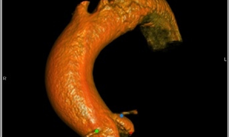
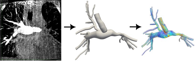
Biomedical and Mechanical Engineering Professor Debanjan Mukherjee is among the researchers working to develop a computer-simulated model that shows the dynamics of a CHD patient's blood flow using 3D Rotational Angiography (RA) images.
"These patients are amongst the most vulnerable and sensitive populations," Mukherjee said. "Even small advancements in their care and treatment outcomes can go a long way in improving their quality of life as they grow older. This remains the dream for our team."
The project, titled "Computational fluid dynamic assessment of patients with congenital heart disease from 3D rotational angiography obtained in the catheterization laboratory," was recently awarded a Colorado Clinical and Translational Sciences Institute Pilot Grant in collaboration with Dr. Jenny Zablah at Children's Hospital of Colorado.
The focus of the research, the 3D RA, is a catheter-based local angiogram, or scan of blood flow through veins and arteries, that provides real-time pressure and flow measurements alongside imaging. It is a highly beneficial yet substantially underexplored imaging option. "Development of high-resolution, in-silico models will enable surgeons to perform hemodynamic analysis prior to a surgical procedure for CHD," Mukherjee said. "The models will facilitate optimal surgical planning and identify indications for interventions."
Credit: University of Colorado at Boulder
Advancing these computer-simulated models will also eliminate the need for additional imaging such as CT or MRI scans, which can be expensive and time-consuming. Mukherjee said those procedures can also introduce these young patients to more radiation and contrast.
The new research will eliminate those additional imaging complexities and facilitate rapid, pre-surgical planning for future pediatric CHD patients.
The team will also address a lack of proper validation in past computer-simulated models. They plan to integrate the computerized blood flow model with space-time varying flow data from 4D flow MRI imaging to validate their predictions. "We remain enthusiastic at the prospect of translating our engineering-based modeling approaches to the realm of patient care," Mukherjee said. "The role that our work, when translated into clinical care, can play for the lives of these children, is the central driving force for all of us."
Source: University of Colorado at Boulder
12.03.2022