© H_Ko – stock.adobe.com
Article • Liver, renal, pancreas, paediatric and multi-organ transplants
Important role for ultrasound in transplantation imaging
Ultrasound plays a pivotal role in the assessment of organ transplant patients. It enables physicians to safely and easily assess progress, identify complications and resolve problems, as well as deliver long-term monitoring. The value of ultrasound in the transplant space was highlighted in a session at ECR 2024, covering liver, renal, pancreas, paediatric and multi-organ transplants with clinicians discussing how it enables them to offer better care for their patients.
Report: Mark Nicholls
Session co-chair Paul Sidhu, Professor of Imaging Sciences at King’s College London, said that in the 1980s, before the much wider use of ultrasound, physicians had little to guide them if a problem arose with a transplant such as with an abnormal liver test function, clotting, thrombosis, or organ rejection.
‘But the introduction of ultrasound transformed how they managed patients,’ he said. ‘They could then clearly move into a different direction – immunosuppressant therapy, angiography, or revascularization.’ The expert argued that, while ultrasound imaging has become a vital part of transplantation, it should be performed more still, and everyone should have the knowlegde of how to do this.
Detecting complications
Dr Gibran Timothy Yusuf from King’s College Hospital NHS Foundation Trust – one of Europe’s biggest transplant centres, conducting 200 liver transplants a year – highlighted how ultrasound in liver transplantation offers an understanding of the anatomy and potential complications, including the hepatic artery and vein, the portal vein and biliary duct.
‘Ultrasound lends itself very well to that evaluation,’ he said. ‘One of the best ways to look at how to use ultrasound is by thinking what the complications can be and how we can potentially detect them.’ This includes vascular complications including formations of pseudoaneurysm, stenosis or occlusion of major vasculature, stenosis of bile ducts, reflux and microlevel ischaemia angiopathy. ‘Ultrasound has a role in the early follow-up, and then also longer term with rejection or recurrent disease,’ he added.
Yusuf also outlined the importance of knowing when to use other modalities for an accurate imaging assessment, particularly when needing to view a wider area. However, he pointed to the clear benefits of ultrasound; it is radiation free, has bedside use, is repeatable, accurate, safe and can be conducted in real time with uses including angiographic assessment, micro- and macrovascular perfusion dynamics.
He said: ‘Multiparametric ultrasound provides comprehensive liver assessment, and the role of B mode and Doppler is critical. CEUS is useful for problem solving, particularly in the early follow-up period. There is a developing role with sheer wave elastography, though we must also acknowledge that complimentary imaging may be needed.’
Paediatric transplants: a wholly different ballgame

Dr Annamaria Deganello, a consultant radiologist at King’s College Hospital since 2013, discussed paediatric liver and multi-visceral transplantation. She focused on the main surgical techniques, post-surgical complications and the role of ultrasound in the post-operative monitoring of paediatric transplants. She said: ‘There is a major difference between paediatric and adult transplantation and the grafts that children receive are very different from the grafts used in adults.’
Deganello said ultrasound, specifically Doppler, remains the most powerful tool to follow these patients up. ‘CEUS microvascular imaging can be used in doubtful cases, especially because you are dealing with tiny vessels,’ she added. ‘But what is really beneficial is that when you are scanning patients, the parents are with you, and you can reassure them immediately rather than waiting for a CT.’
“An excellent problem solver” for renal transplantation
Professor Thomas Fischer, Head of the Ultrasound Centre at Charité in Berlin, concentrated on the monitoring strategy of the early phase after kidney transplantation using MPUS, the benefits of elastography, new broadband Doppler techniques, and CEUS in the early phase after transplantation. He said that ultrasound is the most common modality for assessing kidney transplants and has a role in the post-operative phase from the first 48 hours through to longer term follow-up and is ‘an excellent problem solver’.
Dr Jose Angel Jimenez Lasanta from Hospital Vall d‘Hebron in Barcelona discussed ultrasound in the realms of combined kidney and pancreas transplantation. He discussed the role of ultrasound, including Doppler, in assessing renal/pancreas transplants and indicted when to turn to other imaging modalities for assessment.
Profiles:
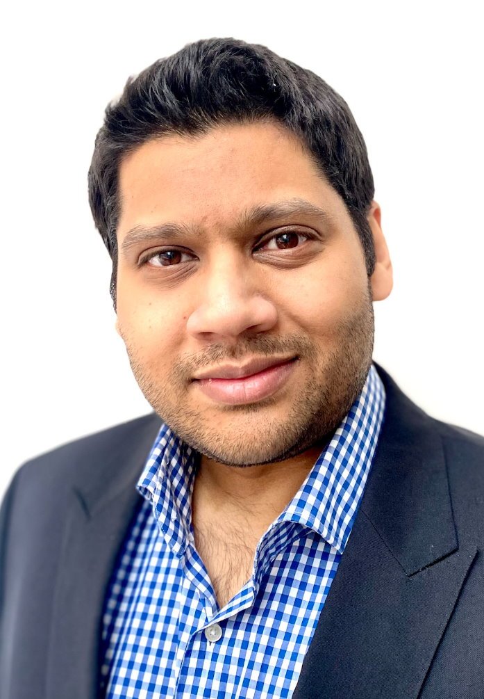
Dr Gibran Timothy Yusuf is Consultant Interventional Radiologist at King’s College Hospital NHS Foundation Trust. He also has a specialist interest in abdominal imaging and ultrasound, including advanced techniques such as contrast enhanced ultrasound, elastography and fusion for which he regularly publishes and is an invited speaker.
Dr Annamaria Deganello is a consultant radiologist at King’s College Hospital, having qualified in 2004 from the University of Padua in Italy before undertaking specialist radiology training. Her specialist interest is in paediatric imaging of complex surgical cases, trauma and hepatobiliary diseases and liver transplantation.
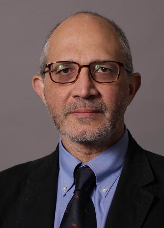
Paul Sidhu is Professor of Imaging Sciences at King’s College London and a Consultant Radiologist in the Department of Radiology at King’ College Hospital. He has published extensively on many aspects of ultrasound and pioneered the introduction of contrast-enhanced ultrasound in the UK. He is Past-President of the European Federation of Societies in Medicine and Biology and the British Medical Ultrasound Society.
16.09.2024