Image source: Kung M et al., BMJ Health & Care Informatics 2024 (CC BY-NC 4.0)
News • Infrared thermography analysis
AI predicts coronary artery disease from facial thermal imaging
Non-invasive, real-time approach is more effective than conventional methods. Testing now required on larger and more diverse numbers of patients, say researchers.
A combination of facial thermal imaging and artificial intelligence (AI) can accurately predict the presence of coronary artery disease (CAD), finds research published in the open access journal BMJ Health & Care Informatics. This non-invasive real-time approach is more effective than conventional methods and could be adopted for clinical practice to improve the accuracy of diagnosis and workflow, pending testing on larger and more ethnically diverse numbers of patients, suggest the researchers.
Current guidelines for the diagnosis of coronary heart disease rely on probability assessment of risk factors which aren’t always very accurate or widely applicable, say the researchers. And while these can be supplemented with other diagnostics, such as ECG readings, angiograms, and blood tests, these are often time consuming and invasive, they add.
Thermal imaging, which captures temperature distribution and variations on the object’s surface by detecting the infrared radiation emitted by that object, is non-invasive. And it has emerged as a promising tool for disease assessment as it can identify areas of abnormal blood circulation and inflammation from skin temperature patterns.
Recommended article

Article • Technology overview
Artificial intelligence (AI) in healthcare
With the help of artificial intelligence, computers are to simulate human thought processes. Machine learning is intended to support almost all medical specialties. But what is going on inside an AI algorithm, what are its decisions based on? Can you even entrust a medical diagnosis to a machine? Clarifying these questions remains a central aspect of AI research and development.
The advent of machine learning technology (AI), with its capacity to extract, process, and integrate complex information, might enhance the accuracy and effectiveness of thermal imaging diagnostics. The researchers therefore set out to look into the feasibility of using thermal imaging plus AI to accurately predict the presence of coronary artery disease without the need for invasive, time consuming techniques in 460 people with suspected heart disease.Their average age was 58; 126 (27.5%) of them were women. Thermal images of their faces were captured before confirmatory examinations to develop and validate an AI assisted imaging model for detecting coronary artery disease.
In all, 322 participants (70%) were confirmed to have coronary artery disease. These people tended to be older and they were more likely to be men. They were also more likely to have lifestyle, clinical, and biochemical risk factors, as well as higher use of preventive meds. The thermal imaging plus AI approach was around 13% better at predicting coronary artery disease than the pre-test risk assessment involving traditional risk factors and clinical signs and symptoms.
As a biophysiological-based health assessment modality, [it] provides disease-relevant Information beyond traditional clinical measures that could enhance [atherosclerotic cardiovascular disease] and related chronic condition assessment
Minghui Kung et al.
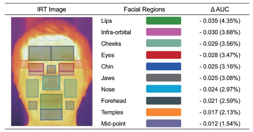
Among the three most significant predictive thermal indicators, the most influential was the overall left-right temperature difference of the face, followed by the maximal facial temperature, and average facial temperature. And, specifically, the average temperature of the left jaw region was the strongest predictive feature, followed by the temperature range of the right eye region and the left-right temperature difference of the left temple regions. The approach also effectively identified traditional risk factors for coronary artery disease: high cholesterol; male sex; smoking; excess weight (BMI); fasting blood glucose, as well as indicators of inflammation.
The researchers acknowledge the relatively small sample size of their study and the fact that it was carried out at only one centre. And the study participants had all been referred for confirmatory tests for suspected heart disease. But they nevertheless write: “The feasibility of [thermal imaging] based [coronary artery disease] prediction suggests potential future applications and research opportunities.” They add: “As a biophysiological-based health assessment modality, [it] provides disease-relevant Information beyond traditional clinical measures that could enhance [atherosclerotic cardiovascular disease] and related chronic condition assessment. The non-contact, real-time nature of [it] allows for instant disease assessment at the point of care, which could streamline clinical workflows and save time for important physician–patient decision-making. In addition, it has the potential to enable mass prescreening.” And they conclude: “Our developed [thermal imaging] prediction models, based on advanced [machine learning] technology, have exhibited promising potential compared with the current conventional clinical tools. Further investigations incorporating larger sample sizes and diverse patient populations are needed to validate the external validity and generalisability of the current findings.”
Source: BMJ
05.06.2024


