News • Noninvasive diagnosis in high-risk CAD patients
Photon-counting CT: “bloom” reduction improves heart disease detection
New ultra-high-resolution CT technology enables excellent image quality and accurate diagnosis of coronary artery disease in high-risk patients, a potentially significant benefit for people previously ineligible for noninvasive screening.
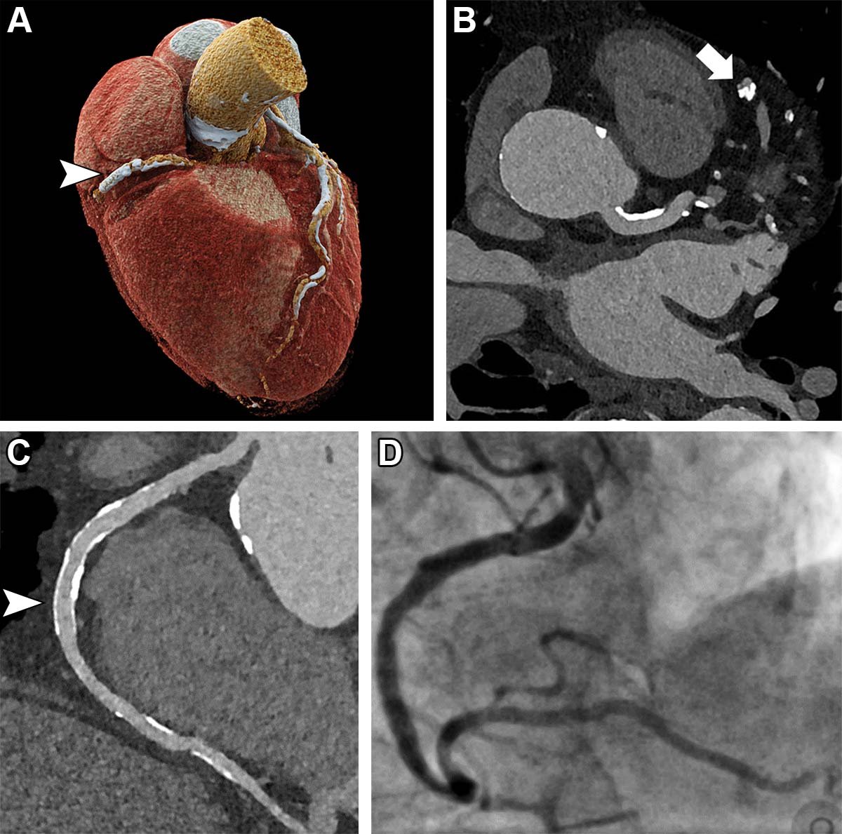
Image source: RSNA
This is according to a study published in Radiology, a journal of the Radiological Society of North America (RSNA).
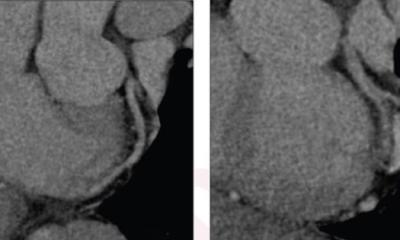
Coronary artery disease is the most common form of heart disease. Coronary CT angiography (CCTA) is highly effective for ruling out coronary artery disease in patients at low or intermediate risk for the disease. Unfortunately, CCTA in a high-risk population is difficult due to a high prevalence of coronary calcifications and stents. Coronary calcifications tend to “bloom” on CCTA, making them appear more extensive than they really are. This results in overestimation of blockages and plaque and false-positive results.
“Consequently, patients may undergo unnecessary, often invasive, testing,” said study lead author Muhammad T. Hagar, M.D., from the Department of Diagnostic and Interventional Radiology at the University of Freiburg in Freiburg, Germany. “This is the reason why current guidelines do not recommend using CCTA in high-risk individuals.”
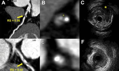
Ultra-high-resolution coronary CT angiography (UHR-CCTA) is a promising tool for the noninvasive assessment of patients at high risk for coronary artery disease. Because it uses recently introduced photon-counting CT scanners, it has not been extensively studied. Dr. Hagar and colleagues compared the diagnostic accuracy of UHR-CCTA with that of the reference standard of invasive coronary angiography (ICA) in 68 patients. The patients had severe aortic valve stenosis, a common, serious valve disease that reduces or blocks blood flow from the heart to the aorta. UHR-CCTA was highly sensitive and specific for coronary artery disease detection. It delivered a median overall image quality score of 1.5 on the 5-point Likert scale, where 1 is excellent and 5 is nondiagnostic. Almost 80% of segments rated as good or excellent.
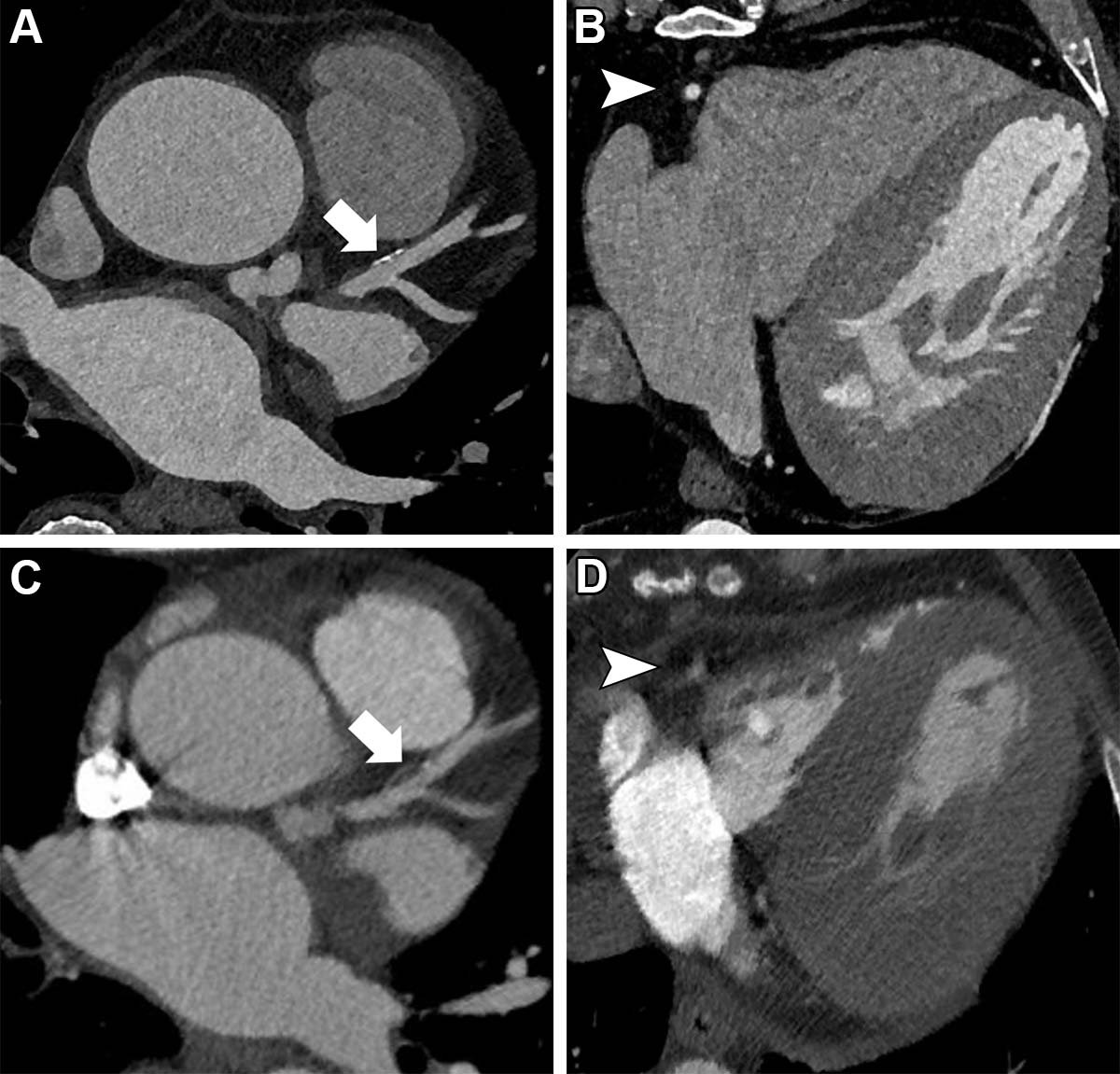
Image source: RSNA
The results suggest that the benefits of noninvasive imaging may soon be available to high-risk patients, Dr. Hagar said. “It appears that the spectrum of patients benefiting from undergoing non-invasive CCTA has been significantly broadened by photon-counting detector technology,” he said. “This is excellent news for these patients and the imaging community.”
The high resolution of UHR-CCTA results from a greater number of emitted photons. This also increases radiation exposure compared with conventional CT scanners. However, Dr. Hagar noted that the technology is at a very early stage and that researchers are developing methods to reduce the amount of radiation exposure. “Currently, the technique is feasible for high-risk patients, in whom the benefits outweigh the risks, but should not be applied to all patients referred for cardiac CT imaging,” he said.
While photon-counting CT is relatively scarce worldwide, experts anticipate that the technology will become more prevalent in the next 10 years [article in German]. “At the University of Freiburg, we had the privilege to work with the technology since its introduction and I am convinced that photon-counting CT is the beginning of a new generation of CT scanners, similar to the introduction to multislice CT 30 years ago,” Dr. Hagar said.
The researchers are exploring the diagnostic ability of photon-counting CT technology in other clinical scenarios such as oncological imaging. In the field of cardiac imaging, they are expanding their research to include subgroups for whom CT imaging is currently not feasible, such as patients with coronary stents. They also are looking into heart muscle assessment with photon-counting CT. Early findings suggest that the technology may improve soft tissue resolution. This would greatly benefit disease characterization. “For me, these are exciting times, and it is just great to be part of a very active group working with this new technology,” Dr. Hagar said.
Source: Radiological Society of North America
21.06.2023