News • MR-Linac measures oxygen levels
One step closer to targeted real time treatment of hypoxic cancers
Imaging researchers have taken a major step towards their ultimate goal of identifying cancers that are starved of oxygen so that altered treatment can be used to target them more effectively.

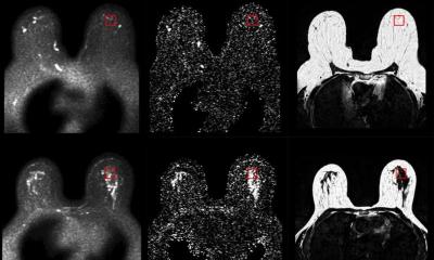
Image source: Dubec et al., Radiotherapy & Oncology 2023 (CC BY 4.0)
The study led by researchers from The University of Manchester, working with scientists at the Institute of Cancer Research, University College London and The University of Leeds, is published in the journal Radiotherapy and Oncology. Funded mainly by the Medical Research Council, the breakthrough was achieved by combining two cutting edge technologies: the research team adapted an MRI scanner that also delivers radiotherapy - called MR-Linac - to be able to also measure oxygen levels in tumours.
Researchers have known since the 1950s that when tumours are starved of oxygen they are difficult to treat effectively, a problem which is particularly true when doctors give radiotherapy. Despite this knowledge, patients with cancer do not get routine tests to evaluate tumour oxygen levels because no single test has been developed that is precise, accurate, cost effective and readily available.
The 11 patients with head and neck cancer in the study, treated at The ChristieNHS Foundation Trust in Manchester, were successfully scanned on the MR-Linac machine and, for the first time, maps of oxygen levels were obtained. However, the technology is relevant to most cancers. The patients first breathed room air through a mask and then pure oxygen to bathe the tumour with the gas. Parts of the cancer that had good levels of oxygen responded differently to those that were oxygen depleted, so the technique - called 'oxygen-enhanced MRI' - revealed which parts of the tumour were oxygen starved and likely to be resistant to radiotherapy.
This imaging lets us see inside tumours and helps us understand why some people with cancer need an extra boost to get effective treatment
James O’Connor
Lead author Professor James O’Connor is a clinician scientist at The University of Manchester, The Christie and The Institute of Cancer Research. He said: “Though it’s clear more work needs to be done, we’re very excited about the potential this technology has to enable daily monitoring of tumour oxygen and we hope to be at a point soon when the technology will guide cancer doctors in how they can best deliver radiotherapy. This imaging lets us see inside tumours and helps us understand why some people with cancer need an extra boost to get effective treatment. This is an important step towards the goal of changing treatment based on imaging biology.”
First author Michael Dubec from The University of Manchester and The Christie said: “The MR-Linac is an exciting technology that combines highly precise imaging and radiotherapy delivery that allows for real-time imaging. We are tremendously excited about what is the first application in humans of 'oxygen-enhanced MRI', developed as a result of a multi-disciplinary team working across the country which has exciting implications on patient outcomes.”
Source: University of Manchester
19.03.2023