Image source: University of Barcelona
News • Molecular mechanism discovered
Researchers find a way to stop harmful cells after cancer treatment
Researchers from the University of Barcelona describe a mechanism for eliminating harmful cells from cancer treatment
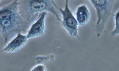
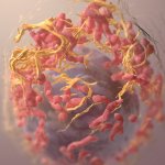
After treating a tumour with chemotherapy and radiotherapy, cells known as senescent cells can appear. These are cells that do not divide, are involved in the ageing process and are resistant to cell death, but are still metabolically active in the human body. When they accumulate, they can jeopardize the patients’ recovery. Now, a study led by researchers from the University of Barcelona (UB) describes for the first time a molecular mechanism that could drive the design of strategies to eliminate senescent cells in cancer patients.
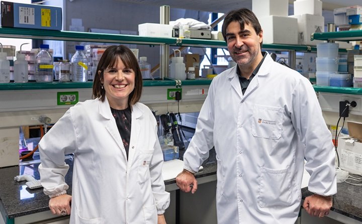
The study, published in the journal Cell Death and Differentiation, from the Nature group, is led by Professor Joan Montero, from the Faculty of Medicine and Health Sciences of the University of Barcelona, and its first author is the researcher Clara Alcon. The study is based on the analysis of human senescent cells in a specific tumour model — melanoma, which affects skin cells known as melanocytes — and, more specifically, melanocytes exposed to chemotherapy or radiation.
During cancer treatment, [...] senescent cells generated by chemotherapy or radiotherapy can survive and regenerate the tumour again or cause premature ageing in patients
Joan Montero
Senescent cells can be caused by various mechanisms “such as exposure to chemotherapy and radiotherapy to treat a tumour, as well as the accumulation of cell damage due to ageing”, says UB professor Joan Montero, a former member of the Institute for Bioengineering of Catalonia (IBEC) and the Networking Biomedical Research Centre on Bioengineering, Biomaterials and Nanomedicine (CIBER-BBN). “During cancer treatment, apart from eliminating tumour cells, the senescent cells generated by chemotherapy or radiotherapy can survive and regenerate the tumour again or cause premature ageing in patients”, says Professor Joan Montero.
Understanding the survival mechanisms of senescent cells will help open up new therapeutic approaches in the field of cancer control. To find answers, the team has worked with multiple melanoma cell lines, molecular markers and cancer therapies to decipher the decisive role of BCL-2 family proteins in senescent cell survival. This family of proteins is key in the regulation of cell death and is made up of different proteins with different functions: they can promote cell death or inhibit it.
Clara Alcon, co-author of the study and researcher at the UB’s Department of Biomedicine: “Most cancer therapies that are used to fight cancer activate the process of apoptosis — a type of programmed cell death — which is controlled by the BCL-2 family of proteins. Therefore, its activity and regulation is decisive in whether or not tumour cells respond to a given therapy. When a tumour is resistant to a treatment, the cause may be an increased activity of anti-apoptotic proteins of this family that block the cell death process”.
Recommended article
Article • Awareness
Focus on cancer
From solid tumors to metastatic carcinomas and leukemia: cancer is among the most common causes of death. Keep reading for latest developments in early detection, staging, therapy and research.
The study methodology successfully employed a technique called BH3 profiling — developed at Professor Anthony G. Letai’s laboratory at the Dana-Farber Cancer Institute and Harvard Medical School — which the team uses to facilitate precision medicine in the fight against cancer. Most importantly, he found differential expression changes that always led to prosurvival adaptation of senescent cells mediated by BCL-XL, an anti-apoptotic protein that prevents the process of cell death.
The authors note that “when we looked at potential therapeutic strategies, we observed senolytic activity, i.e. the ability to kill senescent cells, by specifically inhibiting the BCL-XL protein through compounds such as A-1331852, navitoclax or the PROTAC (proteolysis-targeted chimera) strategy against BCL-XL DT2216”. In the mechanism, they found that “HRK protein levels — a regulatory protein that inhibits BCL-XL — were reduced when senescence was induced and, as a result, led to increased availability of BCL-XL”. The team adds: “Thus, when HRK protein levels are reduced, BCL-XL is free to activate its survival function: it binds to the pro-apoptotic protein BAK and thus slows down the cell death process. In addition, we identified enhanced binding of BCL-XL and BAK that prevented mitochondrial permeabilization and apoptosis”.
“This is the first time that the molecular basis for the anti-apoptotic adaptation of BCL-XL in senescence has been described. This discovery opens the way to develop new therapies that prevent the down-regulation of the HRK protein or displace the binding of BCL-XL to BAK to be used as senolytics”, say the researchers.
For now, the team wants to drive further research studies to see if these molecular processes can be replicated in other tumour types such as lung cancer. “We want to assess whether this molecular mechanism that we now describe is also present in other tumour types, and whether it is mediated by the interaction of the BCL-2 family proteins themselves or others. We also plan to cover new research to analyse the role of the BCL-2 family in the ageing process of different organs and tissues”, conclude Joan Montero and Clara Alcon.
Source: University of Barcelona
26.01.2025


