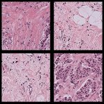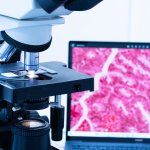
News • Stain-Free Histology
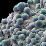
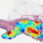
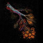
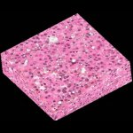
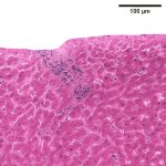
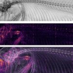
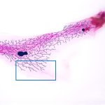
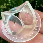
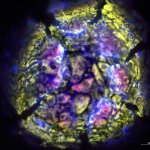
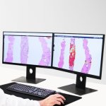
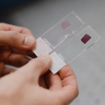
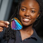
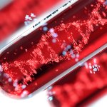
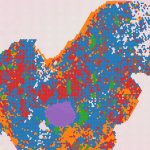
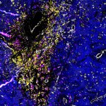
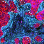
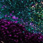
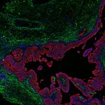
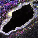
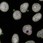
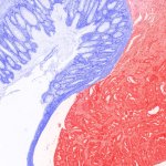
Nanophotonic slides add color to tissue samples
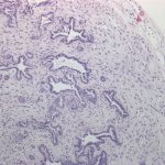
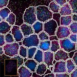
Researchers have developed a method to enhance the cellular features in pathology slides based on “structural color” – the same effect that gives peacock feathers their unique sheen.