© samunella – stock.adobe.com
Interview • AI, modern mammography, and more
Breast imaging breakthroughs presented at EUSOBI 2024
Minimally invasive surgical interventions, innovative imaging and the use of AI: At the upcoming EUSOBI congress in Lisbon, experts present and discuss the latest advances in breast imaging.
Interview: Wolfgang Behrends
We spoke with Tanja Brycker, Vice President, Strategic Development, Breast & Skeletal Health and Gynecological Surgical Solutions at Hologic, ahead of the event about new trends in women’s health, the company’s investment in innovation and education, and what the future of mammography looks like with the rise in AI.
HiE: What are the most significant trends in women’s health that you’ve observed recently, particularly in diagnostic imaging, and how will these be highlighted at the congress?
Tanja Brycker: ‘Minimally invasive surgical interventions for breast cancer, artificial intelligence (AI) and contrast-enhanced mammography (CEM) and biopsy (CEBx) are amongst the most significant current trends in women’s health. The increasing focus on minimally invasive surgical interventions for breast cancer is driven by the potential to improve patient outcomes and well-being, while also benefiting healthcare facilities through reduced demand on resources and a potential reduction in costs.
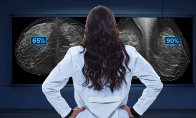
Diagnostic imaging is also no different from other healthcare fields, with AI continuing to be a hot topic, despite having been in use for over a decade. While it is used to manage and clarify diagnostic images, the impact of AI also extends to driving workload efficiencies and the potential to support more personalized medicine. Research has already shown that the next generation of deep-learning technology, such as Hologic’s Genius AI Detection solution, can help significantly enhance performance, surpassing traditional machine learning Computer-Aided Detection (CAD) algorithms in specificity and overall effectiveness.1
Recommended article

Article • Technology overview
Artificial intelligence (AI) in healthcare
With the help of artificial intelligence, computers are to simulate human thought processes. Machine learning is intended to support almost all medical specialties. But what is going on inside an AI algorithm, what are its decisions based on? Can you even entrust a medical diagnosis to a machine? Clarifying these questions remains a central aspect of AI research and development.
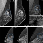
Finally, to mention the growing focus on CEM and CEBx. The synergy between CEM and CEBx has the potential to accelerate cancer diagnosis and workflow efficiencies. CEM pairs 2D and tomosynthesis images, all under one compression, providing anatomical and functional imaging in one exam.2 The contrast agent accumulates where lesions are forming and growing, enabling radiologists to identify it on their 3D mammography systems. When a breast lesion is identified, CEBx enables radiologists to target and acquire tissue samples with the same contrast agent.
These three trends are reflective of the growing awareness of how technology can help address radiologist workload, drive workflow efficiencies and improve patient comfort and satisfaction. By delivering technologies that support diagnostic accuracy, we can improve the experience of everyone involved in the breast diagnostic imaging process.’
What will Hologic be showcasing at the EUSOBI congress?
‘Hologic will highlight a series of innovations designed to help radiologists improve workflow efficiency and diagnostic accuracy – from AI solutions designed specifically for breast cancer diagnosis, to expanded breast biopsy solutions. Attendees can visit Hologic’s booth (M2) at the Lisbon Conference Centre to explore:
- Contrast-enhanced Mammography Solutions that enable clinicians to identify and target lesions that are not visible in 2D or 3D mammography images.
- Hologic's integrated Breast Health AI Software Suite that improves early detection and diagnosis of breast cancer, helps to accelerate workflow, and enables personalized patient care with automated breast density assessment.
- The Brevera® Breast Biopsy System, the world’s first vacuum-assisted breast biopsy solution that integrates tissue acquisition, real-time imaging and verification, and post-biopsy handling. With the newly launched 7-gauge needle, the system now empowers radiology professionals with enhanced clinical versatility to sample larger volume tissue specimens.
Additionally, we will provide a complete medical education programme featuring wide-ranging reading and interventional workshops that provide participants with hands-on experiences. After last year’s successful CEM education workshops, Dr. Jacopo Nori, chief of breast imaging at Careggi University Hospital, will return to lead another series on CEM with his team. He will also be joined by Dr Julia Camps-Herrero, Head of the Radiology Department at University Hospital de la Ribera in Alzira, Valencia, Spain, to discuss new research about using CEM in managing patients with a personal history of breast cancer.
We will also welcome Dr. Sarah Friedewald, associate professor of Radiology and chief of breast imaging at Northwestern Memorial Hospital, for an interactive hands-on reading workshop with our 3DQuorum™ imaging technology. Additionally, Dr. Adnan Duhovic, radiologist at Goldenes Kreuz Private Hospital in Vienna, Austria, will share his experience and learnings for digital breast tomosynthesis (DBT) and the best use of the technology in a nationwide screening programme.’
Recommended article
News • Breast cancer detection
Decade of DBT: 10-year study shows benefits of tomosynthesis
Evidence of the superiority of tomosynthesis for breast cancer detection is stacking up, with new results from a 10-year study further demonstrating the 3D imaging technique's benefits.
Why does Hologic specifically invest in education within the diagnostic imaging sector?
‘At Hologic, we have a longstanding commitment to investment in clinical education. It is essential for radiologists to understand how to correctly use a system for optimized performance and accuracy. CEM is a great example of this, and of the significant investment we have made in an education strategy that supports adoption of, and confidence in, the technology.
We believe that future radiologists should have the opportunity to network with their peers and build relationships with industry leaders, while having access to the latest educational opportunities
Tanja Brycker
Chief of Breast Imaging at Careggi University Hospital, Dr. Nori, has been a great advocate for CEM education, helping EUSOBI attendees learn about CEM image interpretation, its use in pre-surgical planning, and other medical education workshops. We’re proud to work with Dr. Nori on these sessions to build on last year’s activity at our EUSOBI symposium.
Additionally, we continue to sponsor the EUSOBI Breast Imaging Grant for 10 Young EUSOBI Club members to attend the congress as part of our dedication to invest in the future of diagnostic imaging. We believe that future radiologists should have the opportunity to network with their peers and build relationships with industry leaders, while having access to the latest educational opportunities.‘
Can you share any partnerships or collaborations your company has engaged in to foster innovation and education in the field, and will any of these be highlighted at the event?
‘Through our collaboration with Barco, we are excited to announce the arrival of a new monitor for PACS and breast imaging this year. The Coronis Uniti monitor is the biggest and most equipped display system yet available for general radiology and breast imaging. The full-size, 12-million-pixel screen presents images with crystal-clear precision on a large, flexible, bezel-free image area, providing an uninterrupted view of large images, numerous smaller studies, or various combinations and overlays in any layout. EUSOBI attendees will be able to see it on Barco's booth.’
How is AI transforming the field of mammography, and what specific advancements has Hologic made in this area? Will this also be showcased at the EUSOBI congress?
‘AI is revolutionizing mammography by enhancing early detection and diagnostic accuracy, assisting radiologists with identifying subtle abnormalities with greater confidence. As we look at AI today and into the future, it can help empower clinicians to deliver more personalized patient care.
As a global leader in breast health, we see AI playing a major future role in breast imaging. Because of this, we continue to make significant investments in research and innovation related to AI. Our suite of AI solutions is reflective of this, and can help radiologists with optimizing their work, including triaging patients, reading images, and planning for surgery. At our booth at EUSOBI, we’ll showcase this suite of integrated breast health AI solutions for attendees to explore. Most notably, we will introduce attendees to our new Genius™ AI Detection solution – which can help radiologists categorize and prioritize cases by complexity, to optimize workflow and expedite patient care. This new deep-learning AI software is designed to help radiologists detect subtle potential cancers in breast tomosynthesis images. With higher specificity than Hologic’s previous AI solution, the deep-learning software can help detect hard to identify lesions for further examination by the radiologist. Research shows a difference of +9% in observed reader sensitivity for cancer cases using Genius AI Detection technology.3
Also at our booth will be our 3DQuorum imaging technology, which assists radiologists by reducing the number of DBT images they need to review by creating high-resolution, overlapping 6mm "SmartSlices" to expedite reading time. When a radiologist reads SmartSlices instead of 1mm slices, the number of DBT images to review is reduced by two-thirds, leading to an average interpretation time savings of one hour per day.4
AI will continue to revolutionize mammography for years to come and will help improve the radiologist experience. Our suite of integrated AI solutions can help facilitate the workflow efficiency and diagnostic accuracy that they are looking for. We encourage EUSOBI visitors to stop by and learn more.’
References:
- Performance of a traditional machine learning computer-assisted detection (CADe) algorithm versus a deep learning artificial intelligence (AI) algorithm on digital breast tomosynthesis (DBT) studies. Authors: Manisha Bahl (MGH), Constance Lehman (MGH). Accepted as electronic poster.
- Burhenne LJW., Wood SA., D'Orsi CJ., et al. Potential Contribution of Computer-aided Detection to the Sensitivity of Screening Mammography, Radiology 2000; 215:554-562
- Based on analyses that do not control type I error and therefore cannot be generalized to specific comparisons outside this particular study. In this study: The average observed AUC was 0.825 (95% CI: 0.783, 0.867) with CAD and 0.794 (95% CI: 0.748, 0.840) without CAD. The difference in observed AUC was +0.031 (95% CI: 0.012, 0.051). The average observed reader sensitivity for cancer cases was 75.9% with CAD and 66.8% without CAD. The difference in observed sensitivity was +9.0% (99% CI: 6.0%, 12.1%). The average observed recall rate for non-cancer cases was 25.8% with CAD and 23.4% without CAD. The observed difference in negative recall rate was +2.4% (99% CI: 0.7%, 4.2%). The average observed case read-time was 52.0s with CAD and 46.3s without CAD. The observed difference in read-time was 5.7s (95% CI: 4.9s to 6.4s).
- Data on File: Clinical Study Report CSR-00116 Rev. 004
02.10.2024
- AI (865)
- breast cancer (641)
- breast imaging (129)
- education (575)
- mammography (260)
- minimal invasive (206)
- women's health (354)