News • Radiomic parenchymal phenotypes
Dense breasts = high cancer risk? It's not that simple
In one of the larger studies of its kind, researchers have identified six breast texture patterns that may be associated with increased cancer risk, according to a new study.
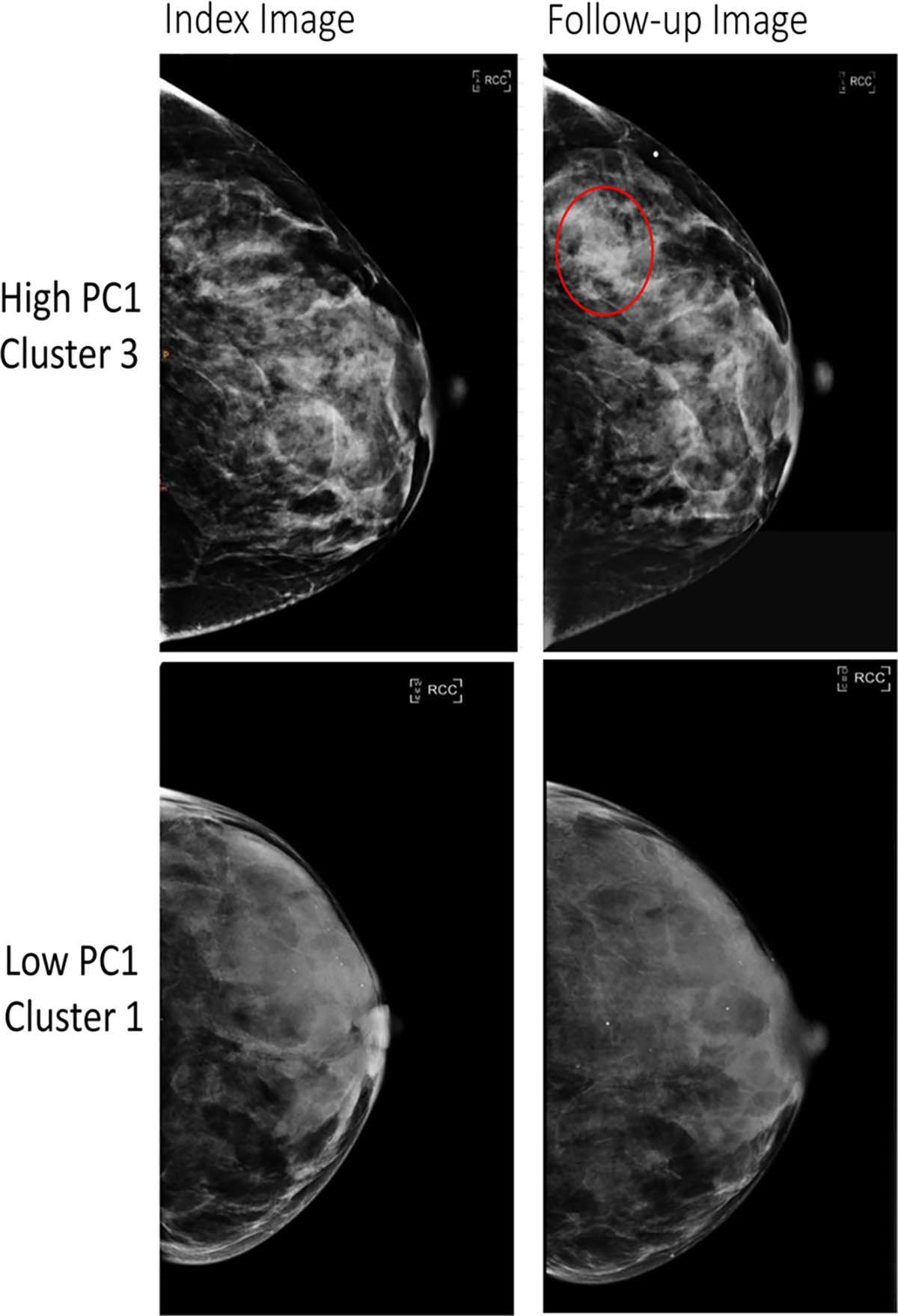
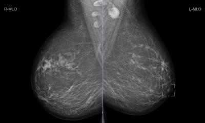
Image credit: Radiological Society of North America (RSNA)
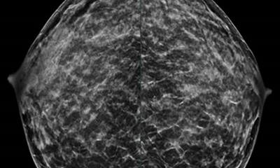
Women with dense breasts, which are breasts with a higher proportion of glandular tissue compared to fatty tissue, make up a large proportion of screening-eligible women. Breast cancer can be difficult to detect on mammograms of dense breasts due to the similarity of appearance of dense breast tissue and cancer growths. Both appear white on the image. Dense breasts are also associated with increased breast cancer risk. Adding to the complexity of the issue is that two women with similar breast density can have substantial differences in their tissue patterns.
Having a greater understanding of the distinct patterns and characteristics of breast tissue, beyond the measurement of breast density, may contribute to improved assessments of breast cancer risk. “We hypothesized that some patterns or phenotypes would be associated with a high risk of future breast cancer and suggest which women may benefit from supplemental screening or prevention strategies,” said one of the senior authors, Celine M. Vachon, Ph.D., a professor of epidemiology at the Mayo Clinic in Rochester, Minnesota. “Other phenotypes could be associated with low risk, ultimately, suggesting less frequent screening.”
The researchers used radiomics on the mammograms of over 30,000 women without a prior history of breast cancer from three different screening cohorts. Radiomics extracts large amounts of quantitative features from medical images to identify patterns and characteristics that might not be visible to the human eye.
Understanding who is at greatest risk of invasive breast cancer, especially the most aggressive types, is crucial for preventing cancer and diagnosing it early for potentially the choice of less intensive treatments
Karla M. Kerlikowske
From the large sample size, the researchers extracted 390 radiomic features that were condensed down into six phenotypes, which are quantifiable characteristics or traits. These six phenotypes were then evaluated on mammograms from over 3,500 women, some who developed breast cancer and some who did not.
Results showed that the radiomic phenotypes were associated with a higher risk of invasive breast cancer in both Black and white women. “We were surprised to find that these radiomic phenotypes showed suggestion of a stronger risk among Black vs. white women,” said co-senior author Despina Kontos, Ph.D., Herbert and Florence Irving Professor of Radiological Sciences and chief research information officer at Columbia University Irving Medical Center. “This is particularly important as breast cancer tends to be more aggressive in Black women, highlighting the need for novel risk factors in this population.”
These phenotypes could also predict a woman’s chances of receiving a false-negative mammogram (when cancer is missed on a mammogram) or an interval cancer diagnosis (cancer diagnosed in between scheduled screenings). “Understanding who is at greatest risk of invasive breast cancer, especially the most aggressive types, is crucial for preventing cancer and diagnosing it early for potentially the choice of less intensive treatments,” said co-senior author Karla M. Kerlikowske, M.D., professor of medicine and epidemiology and biostatistics at University of California San Francisco.
The authors outlined future applications for these phenotypes and how they can complement existing breast cancer risk models. “Our next steps include extending our investigations to larger groups of women in the U.S. population, especially examining 3D mammograms, and combining these radiomic risk factors with genetic and other lifestyle factors to improve our ability to define who is (and who is not) at increased risk of invasive breast cancer,” Dr. Vachon said.
Source: Radiological Society of North America
14.05.2025