Image credit: Canan Dagdeviren/MIT
News • Wearable for monitoring
Breast cancer: In-bra ultrasound scanner to improve early detection
The new device could allow more frequent monitoring of patients at high risk for breast cancer.
When breast cancer is diagnosed in the earliest stages, the survival rate is nearly 100%. However, for tumors detected in later stages, that rate drops to around 25%. In hopes of improving the overall survival rate for breast cancer patients, Researchers at the Massachusetts Institute of Technology (MIT) have designed a wearable ultrasound device that could allow people to detect tumors when they are still in early stages. In particular, it could be valuable for patients at high risk of developing breast cancer in between routine mammograms.
The device is a flexible patch that can be attached to a bra, allowing the wearer to move an ultrasound tracker along the patch and image the breast tissue from different angles. In the new study, the researchers showed that they could obtain ultrasound images with resolution comparable to that of the ultrasound probes used in medical imaging centers. “We changed the form factor of the ultrasound technology so that it can be used in your home. It’s portable and easy to use, and provides real-time, user-friendly monitoring of breast tissue,” says Canan Dagdeviren, an associate professor in MIT’s Media Lab and the senior author of the study.
MIT graduate student Wenya Du, Research Scientist Lin Zhang, Emma Suh, and Dabin Lin, a professor at Xi’an Technological University, are the lead authors of the paper, which appears in Science Advances.
Image credit: Canan Dagdeviren/MIT
For this project, Dagdeviren drew inspiration from her late aunt, Fatma Caliskanoglu, who was diagnosed with late-stage breast cancer at age 49, despite having regular cancer screens, and passed away six months later. At her aunt’s bedside, Dagdeviren, then a postdoc at MIT, drew up a rough schematic of a diagnostic device that could be incorporated into a bra and would allow for more frequent screening of women at high risk for breast cancer.
My goal is to target the people who are most likely to develop interval cancer. With more frequent screening, our goal to increase the survival rate to up to 98%
Canan Dagdeviren
Breast tumors that develop in between regularly scheduled mammograms — known as interval cancers — account for 20-30% of all breast cancer cases, and these tumors tend to be more aggressive than those found during routine scans. “My goal is to target the people who are most likely to develop interval cancer,” says Dagdeviren, whose research group specializes in developing wearable electronic devices that conform to the body. “With more frequent screening, our goal to increase the survival rate to up to 98%.”
To make her vision of a diagnostic bra a reality, Dagdeviren designed a miniaturized ultrasound scanner that could allow the user to perform imaging at any time. This scanner is based on the same kind of ultrasound technology used in medical imaging centers, but incorporates a novel piezoelectric material that allowed the researchers to miniaturize the ultrasound scanner.
To make the device wearable, the researchers designed a flexible, 3D-printed patch, which has honeycomb-like openings. Using magnets, this patch can be attached to a bra that has openings that allow the ultrasound scanner to contact the skin. The ultrasound scanner fits inside a small tracker that can be moved to six different positions, allowing the entire breast to be imaged. The scanner can also be rotated to take images from different angles, and does not require any special expertise to operate.
“This technology provides a fundamental capability in the detection and early diagnosis of breast cancer, which is key to a positive outcome,” says Anantha Chandrakasan, dean of MIT’s School of Engineering, the Vannevar Bush Professor of Electrical Engineering and Computer Science, and one of the authors of the study. “This work will significantly advance ultrasound research and medical device designs, leveraging advances in materials, low-power circuits, AI algorithms, and biomedical systems.”
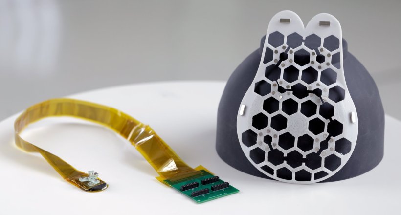
Image credit: Canan Dagdeviren/MIT
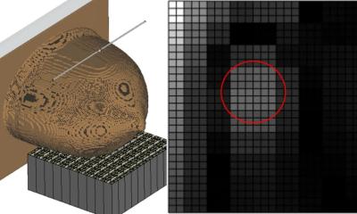
Working with the MIT Center for Clinical and Translational Research, the researchers tested their device on one human subject, a 71-year-old woman with a history of breast cysts. Using the new device, the researchers were able to detect the cysts, which were as small as 0.3 centimeters in diameter — the size of early-stage tumors. They also showed that the device achieved resolution comparable to that of traditional ultrasound, and tissue can be imaged at a depth up to 8 centimeters. “Access to quality and affordable health care is essential for early detection and diagnosis. As a nurse I have witnessed the negative outcomes of a delayed diagnosis. This technology holds the promise of breaking down the many barriers for early breast cancer detection by providing a more reliable, comfortable, and less intimidating diagnostic,” says Catherine Ricciardi, nurse director at MIT’s Center for Clinical and Translational Research and an author of the study.
This conformable ultrasound patch is a highly promising technology as it eliminates the need for women to travel to an imaging center
Tolga Ozmen
To see the ultrasound images, the researchers currently have to connect their scanner to the same kind of ultrasound machine used in imaging centers. However, they are now working on a miniaturized version of the imaging system that would be about the size of a smartphone.
The wearable ultrasound patch can be used over and over, and the researchers envision that it could be used at home by people who are at high risk for breast cancer and could benefit from frequent screening. It could also help diagnose cancer in people who don’t have regular access to screening. “Breast cancer is the most common cancer among women, and it is treatable when detected early,” says Tolga Ozmen, a breast cancer surgeon at Massachusetts General Hospital who is also an author of the study. “One of the main obstacles in imaging and early detection is the commute that the women have to make to an imaging center. This conformable ultrasound patch is a highly promising technology as it eliminates the need for women to travel to an imaging center.”
The researchers hope to develop a workflow so that once data are gathered from a subject, artificial intelligence can be used to analyze how the images change over time, which could offer more accurate diagnostics than relying on the assessment of a radiologist comparing images taken years apart. They also plan to explore adapting the ultrasound technology to scan other parts of the body.
Source: Massachusetts Institute of Technology
29.07.2023