Image source: Modir N, Shahedi M, Dormer J, Ma L, Fei B; Journal of Medical Imaging 2025 (CC BY 4.0)
News • Beyond visible light
Hyperspectral LED endoscope to better detect GI cancers
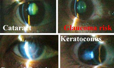
Gastrointestinal cancers remain among the most common forms of cancer. While endoscopy has become a cornerstone of cancer screening and diagnosis over the past two decades, the procedure still misses approximately 8-11% of tumors, due to visibility limitations.
Now, researchers have developed a prototype imaging system that could significantly improve doctors' ability to detect cancerous tissue during endoscopic procedures.
The new approach, reported in the Journal of Medical Imaging, combines light-emitting diodes (LEDs) with hyperspectral imaging technology to create detailed maps of tissue properties that are invisible to conventional endoscopic cameras. Unlike standard endoscopy, which captures images using broad red, green, and blue color channels, hyperspectral imaging records data across numerous narrow wavelength bands, including light beyond the visible spectrum. This allows the system to detect biochemical changes in cancerous tissue that produce distinct spectral signatures.
Our findings suggest that LED-based systems could open new possibilities for hyperspectral imaging applications
Naeeme Modir,
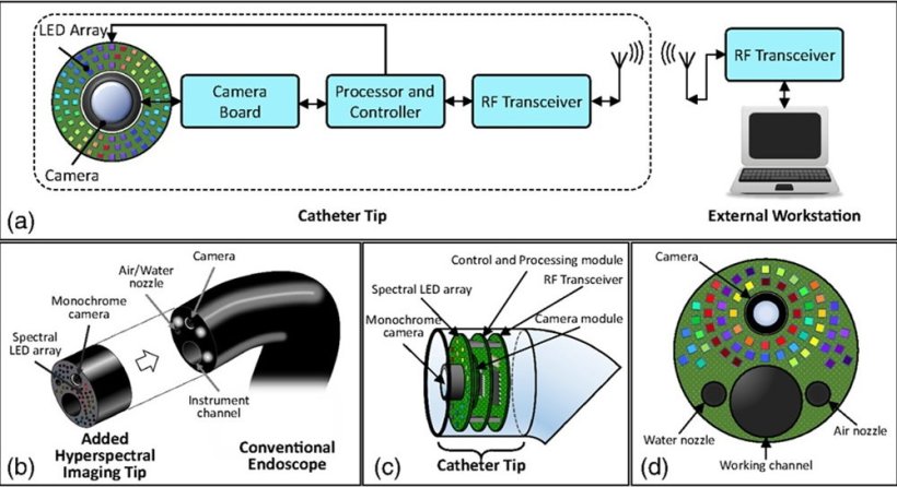
The research team led by Dr. Baowei Fei, Professor and Cecil H. and Ida Green Chair in Systems Biology Science at the University of Texas at Dallas (UT Dallas) Quantitative BioImaging Laboratory, designed and tested a prototype system built around an array of 18 LEDs, each emitting light at different wavelengths ranging from 405 nanometers to 910 nanometers. The system uses a monochrome camera to capture images as each LED illuminates the target tissue in sequence, building up a complete hyperspectral dataset.
The researchers evaluated their LED-based system by imaging both normal and cancerous tissue samples removed during surgery. They investigated how different imaging conditions affected the quality of the hyperspectral data and compared their results with those from a reference hyperspectral camera system used as a gold standard.
The LED-based prototype successfully captured hyperspectral signatures from various tissue types, producing data comparable to the reference system. The researchers found that their approach could achieve imaging rates of over 10 hyperspectral datasets per second, which approaches the real-time speeds needed for practical endoscopic procedures.
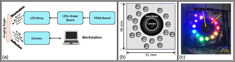
Image source: Modir N, Shahedi M, Dormer J, Ma L, Fei B; Journal of Medical Imaging 2025 (CC BY 4.0)
"Our research shows the feasibility of employing a spectral LED array as the illumination source for high-speed and high-quality hyperspectral imaging," commented Naeeme Modir, PhD candidate at UT Dallas and first author. “Our findings suggest that LED-based systems could open new possibilities for hyperspectral imaging applications,” she remarked.
The LED-based approach offers several potential benefits over existing hyperspectral endoscopy systems. Traditional hyperspectral endoscopes often require fiber optic bundles to deliver light through the working channel of the endoscope, which limits the available space for other medical instruments. The new design places the LEDs directly at the tip of the endoscope, leaving the working channel free for surgical tools or other procedures.
Source: SPIE
11.07.2025