© luchschenF – stock.adobe.com
News • Promising prototype
A “diagnostic wand” to detect oral cancer
A prototype of a new oral cancer diagnosis device, developed by the University of Liverpool, has demonstrated promising results during preliminary tests on histopathology specimens.
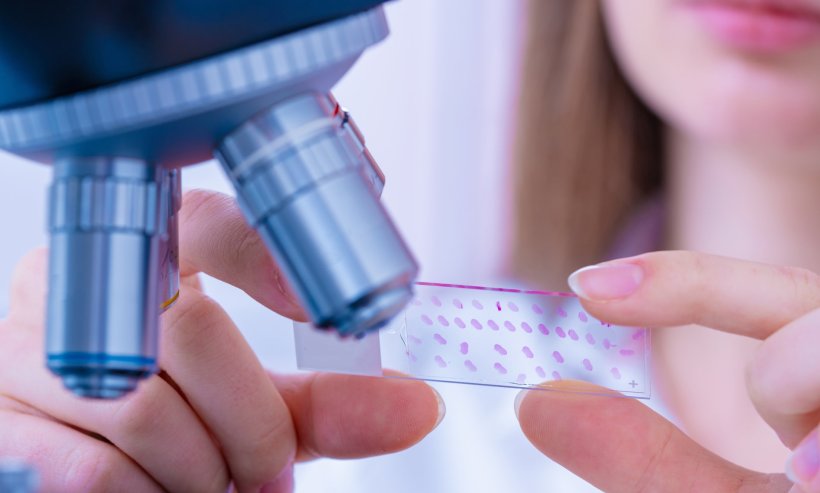
The Liverpool Diagnostic Infrared Wand (LDIR Wand) is being developed by physicists at the University of Liverpool in collaboration with the Liverpool Head and Neck Centre (LHNC). Although still in the early stages of development and tested on a limited number of samples, the LDIR Wand was found to be more accurate at predicting the prognosis of oral cancer lesions than the current haematoxylin and eosin staining technique used to test for mouth cancer.
The LDIR Wand uses a patented machine learning algorithm to analyse infrared spectral images of tissues. It aims to give a value for the percentage area of cancer present in a specimen and, by varying a parameter in the software, can adjust the balance between sensitivity and specificity to aid diagnosis.
Whilst our prototype is currently focussed on oral cancer, our approach should be applicable to any cancer where the diagnosis is based on the analysis of biopsies
Peter Weightman
There are more than 12,000 cases of oral cancer in the UK every year and early diagnosis is critical to improving patient outcomes. Current diagnosis and prognosis methods depend on the analysis of biopsies which is both difficult, prone to subjectivity and time consuming.
Professor Peter Weightman, who has led the development of this pioneering technology, said: “The funding from the National Institute of Health and Care Research’s i4i programme and Cancer Research UK has allowed us to develop a prototype of the LDIR Wand for use in histopathology, and it has been shown to be more accurate than current diagnosis methods. We are hoping to establish a commercial arrangement to market the device so that we can take the next step in the development of this prototype. What is clear is that our technology has the potential to save lives and revolutionise the diagnosis, treatment and prognosis of oral cancer and whilst our prototype is currently focussed on oral cancer, our approach should be applicable to any cancer where the diagnosis is based on the analysis of biopsies.”
Professor Richard Shaw from the LHNC said: “This instrument addresses a difficult clinical problem in head and neck cancer diagnosis. We know that earlier diagnosis is key to saving lives, but predicting cancer risk in oral patches is problematic. For some patients we miss a high risk of cancer developing, but for other patients we cannot safely reassure them without better information.”
The construction and development of the LDIR Wand prototype has been supported with £900k of funding from the National Institute of Health and Care Research’s i4i programme and Cancer Research UK.
The Liverpool Diagnostic Infrared Wand is borne out of pioneering technology developed by University of Liverpool physicist Professor Peter Weightman and its origins date back more than a decade. Whilst working on the ALICE accelerator facility, the UK’s only 4th generation light source based at Daresbury, Professor Peter Weightman, from the School of Physical Sciences, realised that the intense source of infrared light (the InfraRed Free Electron Laser) could be used to study cancer.
Having made this discovery breakthrough, Professor Weightman and his team would take their first steps on the technology development journey. Working with clinicians they developed the technology, securing more than £5 million from a number of medical and science funding sources to support the programme. Professor Weightman added: “It is enormously rewarding to see what began as an idea for a new technology being developed for use by clinicians in the lab. The technology development journey is long and challenging, and I have learnt so much along the way. I am so proud of the progress we have made to get to this point and I am excited for the next steps.”
Source: University of Liverpool
15.01.2025


