News • Water exchange detection
New MRI-based method measures tumor malignancy
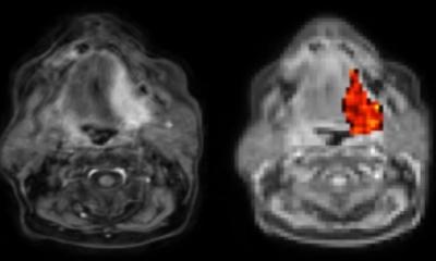
The cycling of water across membrane transporters is an hallmark of the cell metabolism and is potentially of high diagnostic significance for the characterization of tumors and other diseases.
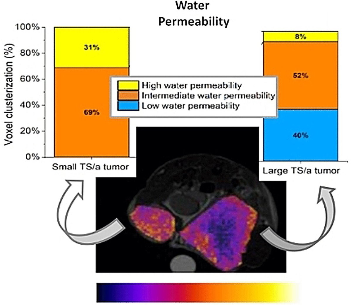
Image source: Di Gregorio et al, Angewandte Chemie 2023; © Wiley-VCH (CC BY-NC-ND 4.0)
In the journal Angewandte Chemie, an Italian research team has now introduced a new MRI-based method for assessing this water exchange. By this method, they were able to estimate the degree of malignancy and the success of treatments in mice tumor models.
Not all cancers are equal. Depending on the type of tumor, a given treatment may be spot on or fail completely. For targeted, effective, treatment that is as gentle as possible, it is important to precisely locate the tumor and determine its malignancy. Magnetic resonance imaging (MRI) provides excellent time- and spatially resolved images for the characterization of tumors. During this procedure, the patient lies in a “tube” in which there is a very strong magnetic field. The spins of protons (the nuclei of hydrogen atoms) align themselves in this magnetic field. Radio waves are beamed in and synchronize the precessions of the spins, temporarily flipping some of them. Depending on the composition of the tissue, this “magnetization” is lost at different times (relaxation). This can be used to compute 3D images. Gadolinium contrast agents reduce the relaxation times. These agents are more concentrated in tumors because their blood vessels are particularly permeable. This increases the contrast and makes it easier to define the tumor.
Contrast agents only spread through the extracellular compartments of the tumor; they do not enter tumor cells. A team led by Giuseppe Ferrauto and Silvio Aime wanted to exploit this feature to determine the degree of water exchange through the cell membrane. Tumor cells are more metabolically active than healthy cells and have more transport proteins and channels in their cell membranes. These proteins also allow water to enter and exit the cell, and the degree of water exchange is a measure of the aggressiveness of a tumor. Yet, classic MRI cannot show this.
Recommended article

Article • Focus on radiology
Magnetic resonance imaging (MRI)
Imaging without ionising radiation: MRI uses magnetic fields to look inside the body. Keep up-to-date with the latest research news, medical applications, and background information on MR imaging.
The team from the University of Torino and IRCCS SDN SynLab in Naples decided to work with a new MRI method called CEST (Chemical Exchange Saturation Transfer). There is constant proton exchange between free water and hydrogen-containing groups in biomolecules, such as the amine groups in creatine. The radio frequencies at which a proton can be “magnetized” depends on the chemical environment of that proton, so frequencies are different for protons in free water and those bound to creatine, for example. With a matching pulse, the creatine-bound protons can be saturated. These protons are exchanged and bind to nearby free water. They keep their “saturated magnetization state” as they do this. If radio waves with the right frequency for free water protons are then pulsed, an increasing number of these protons are already magnetized and cannot absorb the energy (the CEST signal in MR images). Absorption decreases until the proton exchange reaches equilibrium. This makes it possible to draw conclusions about the concentration of creatine and other proton exchanging molecules in a cell, which can be used for cancer phenotyping.
If a contrast agent is then administered and enters the extracellular compartment, the magnetization of the water protons there decreases significantly faster. Because water is exchanged through the membrane, the number of magnetized water protons within the cells also decreases more quickly. This in turn changes the CEST signals. The changes after addition of contrast agent reflect the permeability of the tumor cell membrane to water.
The team tested this method in mouse models for breast cancer with different degrees of malignancy. As expected, the observable water exchange increase as the tumors grew more aggressive. Within the tumors, it was also possible to differentiate between areas of differing malignancy. The cytostatic drug Doxorubicin immediately reduced the water permeability. Hence, the developed method sheds light into tumor phenotype and provides a new tool to assess the outcome of chemotherapy.
Source: Wiley
08.01.2024