Image source: University of Nottingham; adapted from: La Cavera III S, Communications biology 2024 (CC BY 4.0)
News • Novel probe assesses cell stiffness
A 3D endo-microscope for early cancer diagnosis
Researchers at the University of Nottingham have created an endoscopic device that can 3D image the stiffness of individual biological cells and complex organisms, a discovery that could help doctors discover and treat cancer earlier.
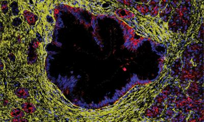
In its early stages, cancer cells are, far softer than normal cells. This allows them to squeeze through tight gaps and rapidly spread throughout the body – known as metastasis. During this process, collections of cells modify their surrounding environment to create stiff tumours that protect them from outside threats. Published in Nature Communications Biology, the new technology can measure the stiffness of these individual cells with a hair-thin endoscopic probe, meaning it will be possible to perform histology (i.e. investigating microscopic cellular tissue) based on abnormal stiffness at the single cell level inside the human body for the first time.
Our device makes it possible to ‘feel for a stiff lump,’ but on a single cellular scale, meaning we could catch cancer early at microscopic cell scales rather than large malignant tumour scale
Salvatore La Cavera III
Dr Salvatore La Cavera III, lead-author and Nottingham Research Fellow in the Optics and Photonics Group at the University of Nottingham, said: “We aim to develop new endoscopic technologies that make diagnostics faster, safer, and clearer for both patients and clinicians. Typically, histopathology requires destructive, invasive biopsies that are not only uncomfortable and potentially damaging for the patient, but require significant logistics such as chemical processing, transportation, and analysis. Our device makes it possible to ‘feel for a stiff lump,’ but on a single cellular scale, meaning we could catch cancer early at microscopic cell scales rather than large malignant tumour scale. It is non-invasive, non-toxic, and very promisingly, is related to technology that can quantitively determine the presence of cancer cells using artificial intelligence – providing a chronically understaffed area with a much-needed solution to a real-world problem that the industry has faced for decades.”
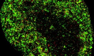
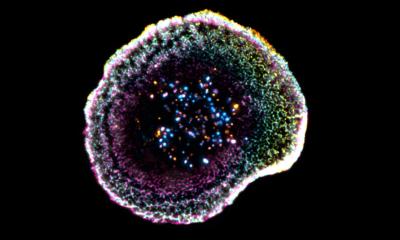
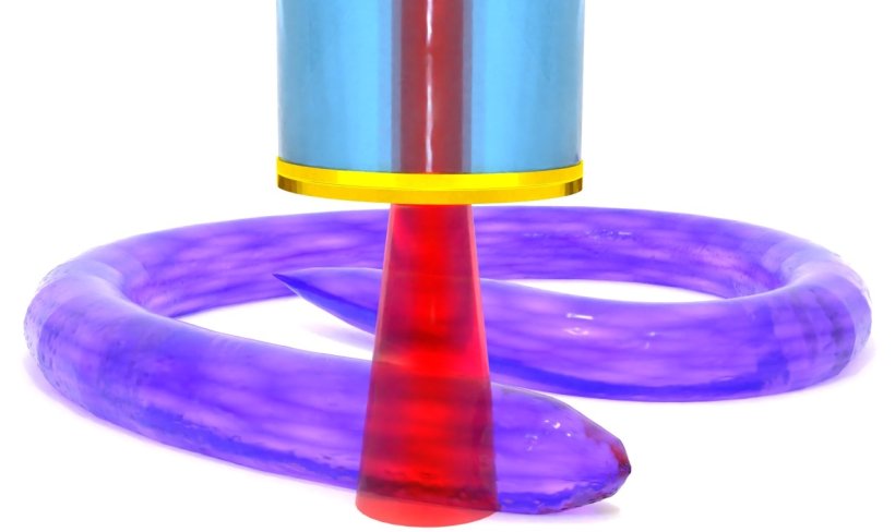
The tool achieves incredibly high imaging resolution, as it can detect the stiffness of objects down to billionths of a metre (nanometres), through a physical phenomenon called Brillouin scattering, where a laser beam interacts with the natural stiffness of a specimen. The team used this to help biologists visualise the 3D stiffness of a microscopic organism, called Caenorhabditis elegans - a free-living worm, known scientifically as a nematode. They were able to provide visualisation and material information about an elusive part of the organism’s anatomy, the cuticle, that up until now had only been imaged through electron microscopes in non-living conditions.
Dr Veeren Chauhan, co-author and Assistant Professor in Whole Organism Analytics at the School of Pharmacy at the University of Nottingham, said: “We have demonstrated the capabilities of this exciting next-generation technology to reveal the physical surface properties of a fully formed microscopic organism in unprecedented detail. Nematodes are an excellent model for human biology and are considered to be the most completely understood animal on the planet in terms of genetics, neurology and developmental biology.
“Until now, these microscopic observations have not been possible in living systems in their natural state with traditional technologies, which typically require animals to be sacrificed. This new probe is significant because it has the potential to allow researchers like us to follow the development of an individual nematodes physical surface properties throughout its entire life cycle, from egg to adult, in approximately three days, which would take upwards of 18 years when considering human development. Therefore, this capability will not only enhance our understanding of nematode biology, but also builds a foundational model for translating these findings to complex biological systems, including human physiology, paving the way for advancements in both basic biological research and clinical diagnostics.”
Source: University of Nottingham
18.04.2024