News • Operator-independent breast cancer screening
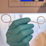
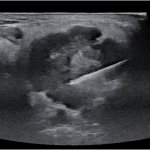
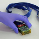
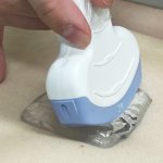
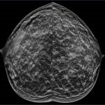
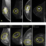
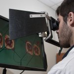
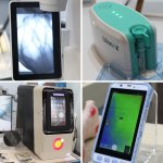
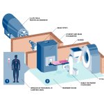
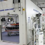
Portable ultrasound system takes the hard part out of breast imaging
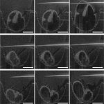
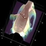
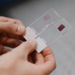
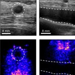
A new portable ultrasound system could make breast imaging more accessible. The device generates high-res, 3D images of breast tissue, requires no expertise to operate and could be used at home.