News • Device biocompatibility research
Improving the body's tolerance to prosthetic implants
An international research team, including scientists from the Institut de Neurociències at the Universitat Autònoma de Barcelona (UAB), has developed a new solution to reduce the immune response triggered by neural prosthetics used after limb amputations or severe nerve injuries.
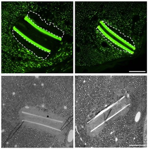
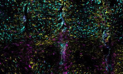
Image source: Turin G, Crugeiras J, Bisquoli C et al., Advanced Healthcare Materials 2025 (CC BY 4.0)
The approach consists of coating the electronic implants (which connect the prosthetic device to the patient’s nervous system) with a potent anti-inflammatory drug. This coating helps the body better tolerate the implant, improving its long-term performance and stability.
Neural electrode implants are commonly used in prosthetics to restore communication between the device and the nervous system. However, their long-term effectiveness can be compromised by the body’s natural immune reaction to foreign objects, which leads to the formation of scar tissue around the implant and can impair its function.
Now, a recent study published in Advanced Healthcare Materials by researchers from the Universitat Autònoma de Barcelona, the Università di Ferrara, the University of Freiburg, and Chalmers University of Technology, conducted as part of the European collaborative project BioFINE, reports a novel method to improve the biocompatibility and chronic stability of these electrodes.
The technique involves activating and modifying the surface of polyimide (a material commonly used for implanted electrodes) using a chemical strategy that enables the covalent binding of the anti-inflammatory drug dexamethasone. This innovation allows the drug to be released at the implant site slowly over at least two months, a critical period when the immune system typically mounts its strongest response.
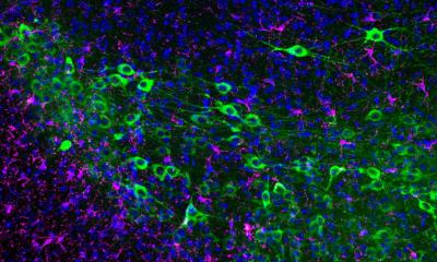
Biological tests showed that this approach reduces inflammation-related signals in immune cells, while maintaining the material’s biocompatibility and mechanical integrity. Animal testing further confirmed that the dexamethasone-releasing implants significantly reduce immune reactions and scar tissue formation around the device.
These findings suggest that the slow and localized release of dexamethasone from the implant surface could extend the functional lifespan of neural prostheses, offering a promising step forward in addressing the long-term challenges of implantable neurotechnology. “This is a main step that has to be complemented by the demonstration in vivo that this coating improves the functional performance of chronically implanted electrodes in the peripheral nerves, for stimulating and recording nerve signals”, says Dr. Xavier Navarro, principal investigator of the UAB team in the BioFINE project.
Source: Universitat Autònoma de Barcelona
03.07.2025