News • Finding fibrosis
Hyperpolarized MRI improves early detection of kidney diseases
Researchers from Aarhus University and Aarhus University Hospital have developed a method that can change the way we diagnose and treat kidney diseases.

Image source: Health AU
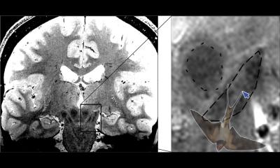
Using an advanced scanner, researchers from Aarhus University, among others, have developed a technology that can detect the earliest changes in the kidney when scar tissue begins to form. "We can measure changes associated with the production of fibrosis earlier than all current methods, which measure the amount of already produced fibrosis," says postdoc Nikolaj Bøgh from the Department of Clinical Medicine at Aarhus University, who is the lead author of a new study on the technology, which was now published in the journal Investigative Radiology.
With the technology, known technically as hyperpolarized 13C-pyruvate MRI, doctors can now see fibrosis formation before it occurs. This makes it possible to start treatment earlier and potentially prevent irreversible damage to the kidney. "The method takes a completely new approach, catching scar tissue early by imaging the building blocks that fibrosis consists of," explains Nikolaj Bøgh.
The scans can open a whole new front in the treatment of kidney patients. We expect to use them to tailor treatment to the individual patient when we can identify patients who need rapid and targeted treatment
Nikolaj Bøgh
The technology works by injecting a special form of pyruvate, a natural substance in the body's energy production, into the patient's body. When pyruvate molecules are hyperpolarized, their magnetic signals are significantly amplified, more than 20,000 times. This allows tracking their conversion in the body using an MRI scanner. By tracking how pyruvate converts into other substances, doctors can detect early signs of fibrosis before there are visible structural changes that can be captured with standard methods.
The method is not only more effective but also safer and more comfortable for patients, as it eliminates the need for invasive biopsies. "The scans can open a whole new front in the treatment of kidney patients. We expect to use them to tailor treatment to the individual patient when we can identify patients who need rapid and targeted treatment," explains Nikolaj Bøgh.
The technology has the potential to be applied in other areas beyond kidney diseases. Fibrogenesis, which the technology measures, is not unique to the kidneys but can also be relevant to other organs, such as the heart in certain types of heart failure. However, to transfer the technology from the laboratory to the clinic, further trials on patients are required.
Nikolaj Bøgh and his colleagues have already initiated three clinical studies on patients with various kidney diseases. The studies aim to demonstrate the method's value, including in identifying diabetic patients at high risk of developing kidney disease. Although the technology shows great potential, it will be challenging to implement it widely in clinical practice. There are only about 24 of these advanced scanners in the world, and even fewer can examine humans with the technology. Over the coming years, however, researchers hope to see the technology become more accessible and widespread. "We hope to see the technology become more available so that patients can benefit from a scanner that is more than 20,000 times more sensitive than the conventional scanners we use in hospitals today," says Nikolaj Bøgh.
Source: Aarhus University
14.07.2024