© sdecoret – stock.adobe.com
News • Gut function and microbiome analysis
MRI reveals impact of gluten free diet on people with celiac disease
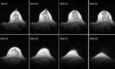
Experts have used magnetic resonance imaging (MRI) to better understand the impact a gluten free diet has on people with celiac (or coeliac) disease, which could be the first step towards finding new ways of treating the condition.
The MARCO study – MAgnetic Resonance Imaging in COliac disease – which is published in Clinical Gastroenterology and Hepatology (CGH), was led by experts from the School of Medicine at the University of Nottingham, alongside colleagues at the Quadram Institute.
Celiac disease is a chronic condition affecting around one person in every 100 in the general population. When people with celiac disease eat gluten, which is found in pasta and bread, their immune system produces an abnormal reaction that inflames and damages the gut tissue and causes symptoms such as abdominal pain and bloating.
The findings increase our understanding of gut function and physiology in celiac disease and open the possibility of developing prebiotic treatments to reverse the negative impact of the gluten free diet on the microbiome
Luca Marciani
The only treatment is a life-long commitment to a gluten free diet, which helps recovery of the gut tissue but still leaves many patients with gastrointestinal symptoms.
Luca Marciani, Professor of Gastrointestinal Imaging at the University, led the study. He said: “Despite being a common chronic condition, we still don’t precisely know how celiac disease affects the basic physiological functioning of the gut and how the gluten free diet treatment may further change this. We launched the MARCO study to try and address this issue, by using MRI along with gut microbiome analysis to give us new insights into how a gluten-free diet affects people with celiac disease.”
The team recruited 36 people who had just been diagnosed with celiac disease and 36 healthy volunteers to participate in the study. Images were taken of their guts with MRI, along with blood and stool samples. The patients then followed a gluten free diet for one year and came back to repeat the study. The healthy participants came back one year later too and repeated the study, but they did not follow any diet treatment. The study found that the newly diagnosed patients with celiac disease had more gut symptoms, more fluid in the small bowel and that the transit of food in the bowel was slower than in the healthy controls.
Recommended article

Article • Human-bacterial symbiosis
The value of the human microbiome
The human species maintains symbiotic relationships with a multitude of microbial organisms that colonise the inside as well as the surface of the body. Scientists, for a long time, underestimated the significance of these organisms for humans.
The microbiota (the ‘bugs’ living in the colon) of the patients showed higher levels of ‘bad bugs’ such as E.coli. After one year of a gluten free diet, gut symptoms, bowel water and gut transit improved in the patients, but without returning to normal values. By contrast, the gluten free diet reduced some of the ‘good bugs’ in the microbiota, such as Bifidobacteria associated with reduced intake of starch and wheat nutrients, due to the different diet.
The patient study was conducted by Radiographer Dr Carolyn Costigan, from Nottingham University Hospitals, as part of her PhD studies at the University of Nottingham. Professor Marciani said: “It was particularly interesting to see how the imaging results on gut function correlated with changes in the ‘bugs’ in the colon microbiota. The findings increase our understanding of gut function and physiology in celiac disease and open the possibility of developing prebiotic treatments to reverse the negative impact of the gluten free diet on the microbiome.”
Dr Frederick Warren from the Quadram Institute said: “This study is the result of an exciting and innovative research collaboration bringing together medical imaging technology and gut microbiome analysis. We provide important insights which pave the way for future studies which may identify novel approaches to alleviate long-term symptoms in celiac patients.”
Source: University of Nottingham
08.12.2024


