© Fabien Kawecki/Inserm
News • Biomaterial-based device
Heart valve made of human collagen opens up new treatment avenues
Researchers from Inserm have developed a pulmonary valve using human collagen. This device could ultimately be a game-changer in the treatment of paediatric heart diseases, such as tetralogy of Fallot.
These findings form part of broader research conducted at BioTis (Inserm/Université de Bordeaux), a tissue bioengineering lab in Bordeaux which develops tissues made from biological material obtained from human cells. This study, published in Science Translational Medicine, opens up new therapeutic avenues in the longer term for young patients with tetralogy of Fallot, for whom the current treatment options continue to cause numerous complications.
We have obtained proof of concept that the valve we have designed is functional and can easily be fitted following the same surgical procedures as in humans, which is promising if we are to move on to clinical studies
Fabien Kawecki
Tetralogy of Fallot is a congenital heart malformation that affects one in 4 000 births. It involves pulmonary stenosis, which is when the outflow tract from the heart’s right ventricle to the pulmonary artery is narrowed. This prevents the normal flow of blood to the lungs, leading to reduced blood oxygen levels. This abnormality can be corrected with surgery to widen the pulmonary pathway to restore normal blood flow. This involves removing the pulmonary valve, and then reconstructing it from either synthetic Teflon membranes or ‘biological’ leaflets made from chemically treated animal tissue.
Both solutions present major drawbacks – the first and foremost being a reaction from the immune system, which seeks to reject these foreign bodies. Together with a chronic inflammatory reaction, this phenomenon can also lead to other complications such as thrombosis and calcification. What is more, the valves made from these materials are prone to the development of bacterial infections. Finally, they are not designed to adapt to the patient’s growth and changing morphology: this means that as the patient ages, other operations will be required to replace the initial valve.
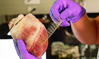
Therefore, the team led by Inserm researcher Fabien Kawecki wanted to find new solutions, and so has developed a ‘new-generation’ biological pulmonary valve, designed from collagen-rich leaflets produced by cells. Collagen is a structural protein highly abundant in the human body, which helps to support many tissues and organs. The researchers thus based themselves on the approach developed over the last decade at BioTis, which consists of culturing human cells in the laboratory to obtain deposits of collagen-rich extracellular matrix.
These collagen deposits form leaflets that can be used to devise, as in this study, pulmonary valves. The major advantage is that since collagen does not vary from one person to another, these entirely biological and chemically undenatured leaflets are not considered by the body as foreign bodies to be rejected.
This digital model and that of the organosynthetic heart could be valuable tools for researchers and surgeons, enabling them in the future to test new biomaterials and medical devices, as well as to train in new surgical approaches before moving to animals and then humans
Fabien Kawecki
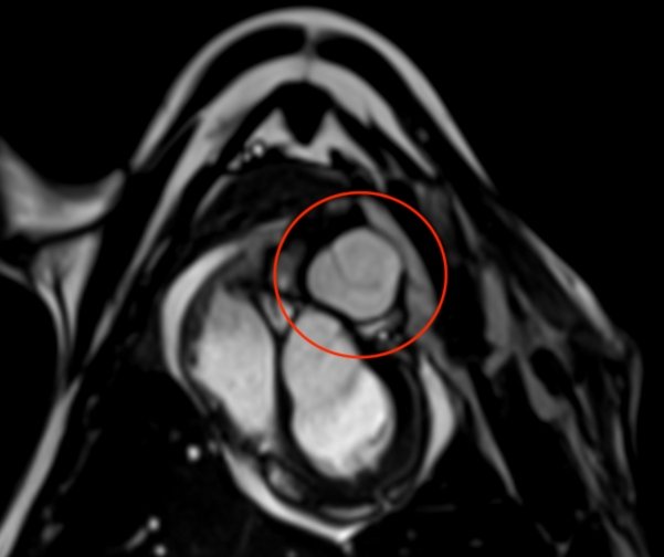
In the study, Kawecki and his team tested the use of their biological leaflets to reconstruct a pulmonary valve in an ‘organosynthetic’ heart model developed by their US collaborators at the Massachusetts Institute of Technology (MIT). It is a bioartificial heart that uses pneumatic muscles to reproduce the functioning of the human heart and to control heart beats. From this model it is possible to gather valuable data on the functionality of the valve. Then, working with cardiac surgeons at Bordeaux University Hospital, the scientists also implanted the valve for seven days in an animal model (sheep), performing the same surgical procedures and using the same tools as those used for this type of operation in humans.
‘Thanks to our two models, we have obtained proof of concept that the valve we have designed is functional and can easily be fitted following the same surgical procedures as in humans, which is promising if we are to move on to clinical studies in a few years’ time. Implanting our valve has restored the direction of blood flow through the pulmonary pathway without generating valve leakage. We also observed that after only 7 days of implantation, there was good integration of the valve with the animal’s native tissue. In addition, we have seen on our valve the presence of smooth muscle cells which will play an important role in its remodelling and growth,’ explains Kawecki.
Using the data collected in both models, the scientists were also able to develop a digital model that will allow the functionality and clinical utility of different biomaterials to be tested before being implanted in animals. ‘This digital model and that of the organosynthetic heart could be valuable tools for researchers and surgeons, enabling them in the future to test new biomaterials and medical devices, as well as to train in new surgical approaches before moving to animals and then humans,‘ explains Kawecki.
For the team, the next step is to implant the valve over longer periods of time (16 weeks, then one year) in animal models, to ensure that it functions correctly over the long term and adapts to the growth of the animal over time. Beyond that, if the results are conclusive, clinical trials could be envisaged. The team has already filed a patent for the use of the lab-designed biomaterial as a pulmonary valve and hopes to test its utility in various cardiovascular diseases in adults and children in the future.
Source: Inserm
17.07.2024