News • Mechanical forces guide axon growth
Magnet-pulling brain cells to treat Parkinson's
A collaborative study led by Professor Vittoria Raffa at the University of Pisa and Assistant Professor Fabian Raudzus at the Center for iPS Cell Research and Application, Kyoto University, has unveiled a novel approach that uses magnetically guided mechanical forces to direct axonal growth, aiming to enhance the effectiveness of stem cell-based therapies for Parkinson's disease (PD) and other neurological conditions.
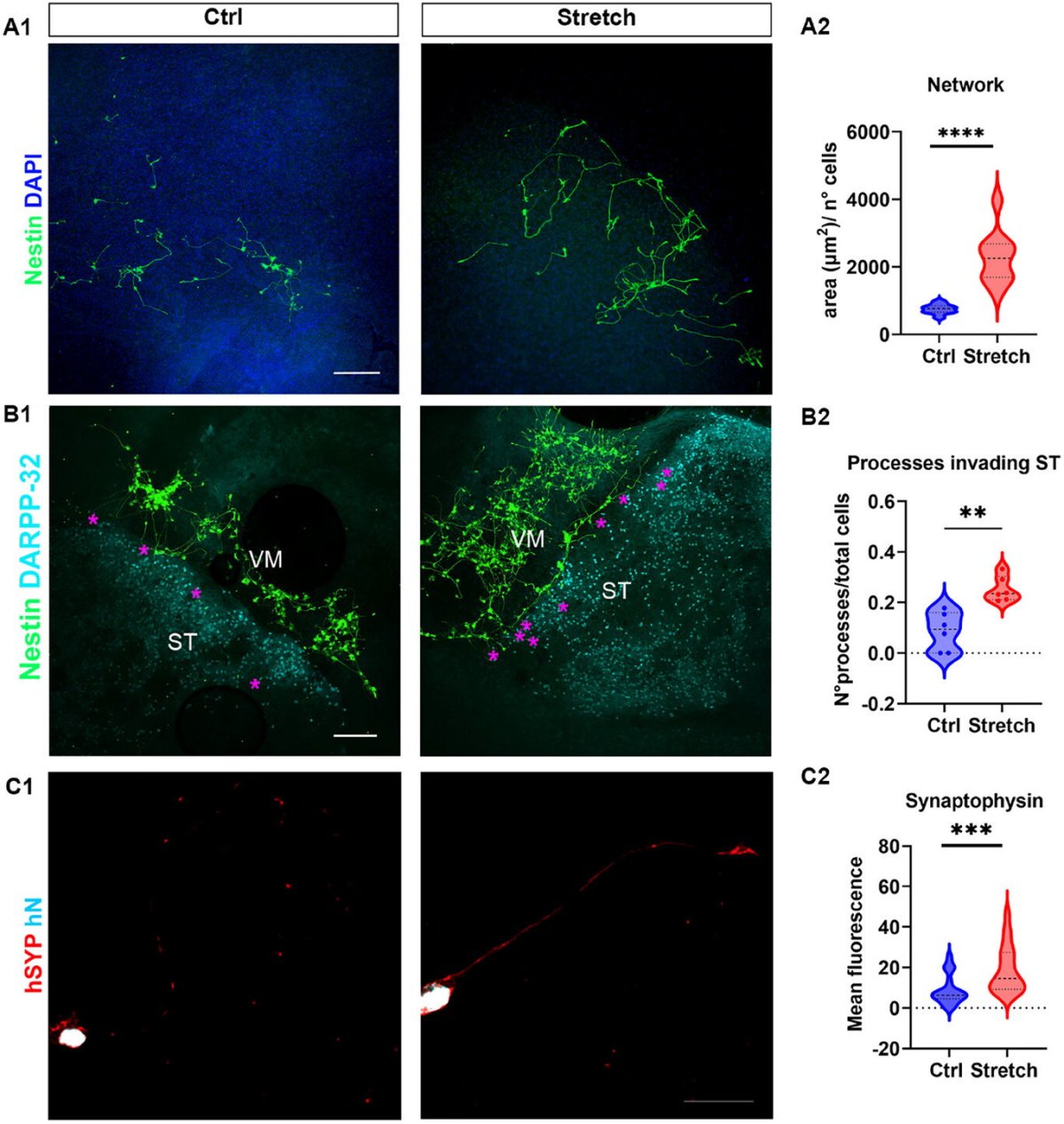
Image source: De Vincentiis S, Capitanini E, Kira K et al., Advanced Science 2025 (CC BY 4.0)
Parkinson's disease is characterized by the progressive degeneration of dopaminergic neurons in the substantia nigra (SN), which project to the striatum (ST) via the nigrostriatal pathway. The loss of these connections leads to dopamine deficiency and the onset of motor symptoms. While cell replacement therapies using human stem cell-derived dopaminergic progenitors have shown encouraging results in clinical trials, a key limitation remains: the inability to guide the axons of transplanted cells over long distances to their appropriate targets in the adult brain.
To address this, the researchers developed a technique—called nano-pulling—to leverage magnetic nanoparticles (MNPs) and external magnetic fields to apply controlled mechanical forces within transplanted neural cells, thereby guiding their axonal extensions to target brain regions. To test this system, the research team first constructed an organotypic brain slice model that mimics early-stage PD by co-culturing brain sections containing SN and ST. They transplanted human neuroepithelial stem (NES) cells, preloaded with MNPs, into the SN region. Upon exposure to a magnetic field, the MNPs generated piconewton-scale forces within the cells, stimulating axonal growth in the direction of the magnetic gradient.
The research was published in the journal Advanced Science.
The study found that nano-pulling significantly enhanced the length and alignment of neural projections toward the striatum. Transplanted cells exhibited increased branching, greater synaptic vesicle formation, and improved microtubule stability—key indicators of neuronal maturation and functional integration. These effects were observed in both NES cells and human iPS cell-derived dopaminergic progenitors, thus demonstrating the versatility and clinical relevance of the approach.
Importantly, the use of magnetic nanoparticles and magnetic fields—both of which are already employed in clinical imaging and therapeutic applications—supports the translational potential of nano-pulling. The researchers also confirmed that the technique did not compromise cell viability or tissue integrity, even after extended periods of mechanical stimulation.
This work represents a significant step forward in the development of regenerative therapies for neurodegenerative diseases. By enabling the reconstruction of the nigrostriatal pathway through guided axon growth, nano-pulling could enhance the efficacy of cell transplantation strategies and help restore functional connectivity in the brain. Future studies will focus on optimizing nanoparticle properties, evaluating long-term outcomes in vivo, and exploring the broader applicability of this technique in other models of central nervous system injury and disease.
Source: Center for iPS Cell Research and Application, Kyoto University
07.07.2025