Image source: Lunghammer - TU Graz
News • New machine learning method
Seeing the beating heart with better MRI videos
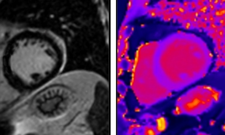
Medical imaging using magnetic resonance imaging (MRI) is very time-consuming since an image has to be compiled from data from many individual measurements. Thanks to the use of machine learning, imaging is also possible with less MRI measurement data, which saves time and costs.
However, the prerequisite for this is perfect images that can be used to train the AI models. Such perfect training images do not exist for certain applications, such as real-time (moving image) MRI, as such images are always somewhat blurred. An international research team led by Martin Uecker and Moritz Blumenthal from the Institute of Biomedical Imaging at Graz University of Technology (TU Graz) has now succeeded in generating precise live MRI images of the beating heart even without such training images and with very little MRI data with the help of smartly trained neural networks. Thanks to these improvements, real-time MRI could be used more frequently in practice in the future.
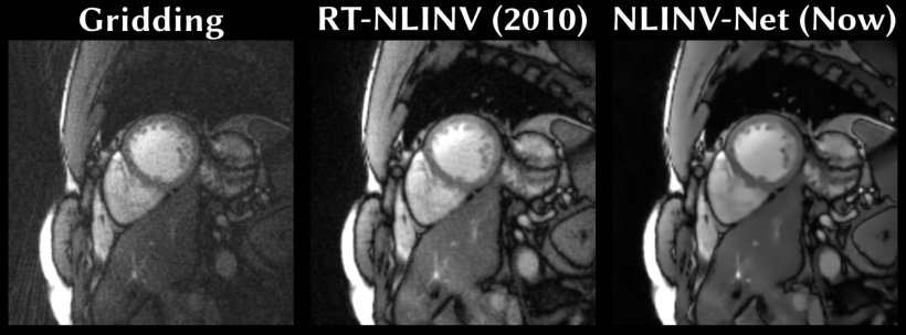
Image source: TU Graz - Institute of Biomedical Imaging
Martin Uecker and Moritz Blumenthal used self-supervised learning methods to train their machine learning model for MRI imaging. The basis for training the model is not pre-curated perfect images, but a subset of the initial data from which the model is to reconstruct the images. Moritz Blumenthal explains it like this: “We divided the measurement data provided by the MRI device into two portions. From the first, larger data portion, our machine learning model reconstructs the image. It then attempts to calculate the second portion of the measurement data withheld from it on the basis of the image.” If the system fails to do this or does so poorly – according to the underlying logic – the previously reconstructed image must have been incorrect. The model is updated, it creates a new improved image variant and attempts to calculate the second data portion again. This process runs for a number of rounds until the result is consistent. In this training process, the system learns from a large number of such reconstructions what good MRI images should look like. Later, during the application, the model can then directly calculate a good image.
Image source: Lunghammer - TU Graz
“Our process is ready for application,” says Martin Uecker, “even if it will probably be a while before it is actually used in practice.” The method can be used for many other MRI applications to make them faster and therefore cheaper. This includes quantitative MRI, for example, in which physical tissue parameters are precisely measured and quantified. “This allows radiologists to access precise data for diagnoses instead of having to interpret images based on differences in brightness using their professional experience,” explains Martin Uecker. “Up to now, however, quantitative MRI measurements have often taken a very long time. With our machine learning model, we were able to speed up these measurements considerably without any loss of quality.”
The research results, which were recently published in the journal Magnetic Resonance in Medicine are the result of an international and interdisciplinary collaboration of the Institute of Biomedical Imaging. Participants included Christina Unterberg (cardiologist at the University Medical Centre Göttingen), Markus Haltmeier (mathematician at the University of Innsbruck), Xiaoqing Wang (MRI researcher at Harvard Medical School) and Chiara Fantinato (Erasmus student from Italy). The algorithms and MRI data are freely available so that other researchers can reproduce the results directly and build on the new method.
Source: Graz University of Technology
30.09.2024