News • Synthetic fibrosis distributions
AI generates ‘scarred hearts’ for better cardiac treatments
A new study demonstrates how artificial intelligence can predict the success of heart procedures without relying solely on real patient data
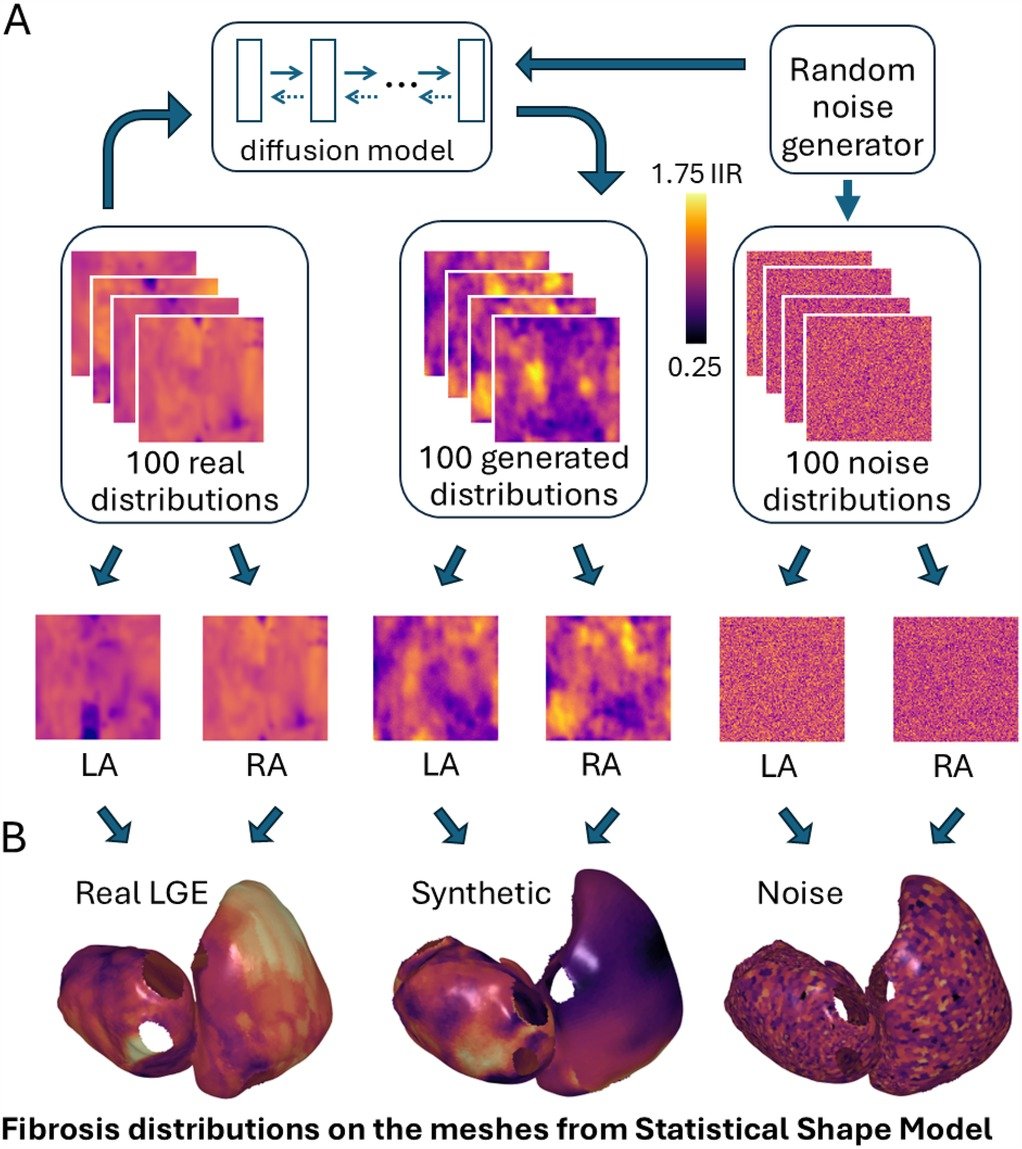
Image source: Zolotarev AM, Johnson K, Mohammad Y et al., Frontiers in Cardiovascular Medicine 2025 (CC BY 4.0)
Researchers from Queen Mary University of London have developed an AI tool that creates synthetic yet medically accurate models of fibrotic heart tissue (heart scarring), aiding treatment planning for atrial fibrillation (AF) patients. The study, published in Frontiers in Cardiovascular Medicine, could lead to more personalised care for patients affected by this common heart rhythm disorder.
Fibrosis refers to scar tissue that develops in the heart, often as a result of ageing, long-term stress or the AF condition itself. These patches of stiff, fibrous tissue disrupt the heart's electrical system potentially causing the irregular heartbeat characteristic of AF. Currently assessed through specialised MRI scans (LGE-MRI), the pattern and distribution of this scarring significantly influences treatment outcomes.
Atrial fibrillation is frequently treated with ablation – a procedure where doctors create small, controlled scars to block erratic electrical signals. However, success rates vary considerably, and predicting which approach will work best for individual patients remains challenging. While AI has shown promise in forecasting outcomes, its development has been hampered by limited access to high-quality patient imaging data.
It's about providing clinicians with a sophisticated simulator – allowing them to test different treatment approaches on a digital model of each patient's unique heart structure before performing the actual procedure
Alexander Zolotarev
“LGE-MRI provides vital information about heart fibrosis, but obtaining enough scans for comprehensive AI training is challenging,” explains first author Dr Alexander Zolotarev of Queen Mary University of London. "We trained an AI model on just 100 real LGE-MRI scans from AF patients. The system then generated 100 additional synthetic fibrosis patterns that accurately mimic real heart scarring. These virtual models were used to simulate how different ablation strategies might perform across varied patient anatomies.”
The team's advanced diffusion model produced synthetic fibrosis distributions that matched real patient data with exceptional accuracy. When these AI-created patterns were applied to 3D heart models and tested against various ablation approaches, the resulting predictions proved nearly as reliable as those using genuine patient data. Crucially, this method protects patient privacy while enabling researchers to study a much broader range of cardiac scenarios than conventional methods allow.
The research highlights AI's emerging role as a clinical support tool rather than a decision-maker. “This isn't about replacing doctors' judgement,” Dr Zolotarev emphasises. “It's about providing clinicians with a sophisticated simulator – allowing them to test different treatment approaches on a digital model of each patient's unique heart structure before performing the actual procedure.”
This work forms part of Dr Caroline Roney's UKRI Future Leaders Fellowship project, which aims to develop personalised 'digital twin' heart models for AF patients. Dr Caroline Roney of Queen Mary University of London, lead author of the study, said: “We’re very excited about this research as it addresses the challenge of limited clinical data for cardiac digital twin models. Our key development enables large scale in silico trials and patient-specific modelling aimed at creating more personalised treatments for atrial fibrillation patients.”
With atrial fibrillation affecting 1.4 million people in UK and ablation failing in half of cases, the technology could significantly reduce repeat procedures. Importantly, the AI approach addresses two critical healthcare challenges: limited patient data availability and the ethical need to protect sensitive medical information.
Source: Queen Mary University of London
15.04.2025