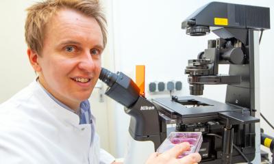
Image source: ACS; photo credit: Tammy T. Chang, MD, PhD
News • Research aboard the International Space Station
How liver tissue from the ISS may transform tissue engineering
Pioneering space research leverages microgravity for tissue engineering breakthroughs
An “out-of-this-world” project has the potential to transform the future of tissue engineering and liver transplantation through innovative research conducted aboard the International Space Station (ISS).
Led by Tammy T. Chang, MD, PhD, FACS, a professor of surgery at the University of California, San Francisco, the Chang Laboratory for Liver Tissue Engineering is pioneering the self-assembly of human liver tissues in low Earth orbit (LEO) — the area of space below an altitude of 1,200 miles. This process could significantly enhance the development of complex tissues for medical use on Earth. Strategies to transport these tissues back to Earth and relevant experimental results were presented at the American College of Surgeons (ACS) Clinical Congress 2024 in San Francisco, California.
Our goal is to develop robust preservation techniques that allow us to bring functional tissues back to Earth, where they can be used for a range of biomedical applications, including disease modeling, drug testing, and eventually, therapeutic implantation
Tammy Chang
This method leverages the unique environment of microgravity to address the limitations of current tissue engineering techniques on Earth. For example, the use of artificial matrices that provide a framework on which cells grow can introduce outside materials and alter cellular function. “Our findings indicate that microgravity conditions enable the development of liver tissues with better differentiation and functionality than those cultured on Earth,” said Dr. Chang. “This represents a critical step toward creating viable liver tissue implants that could serve as an alternative or adjunct to traditional liver transplants.”
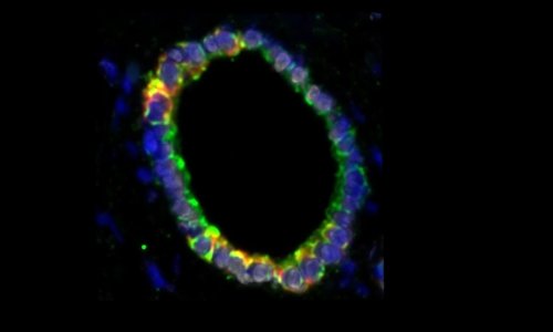
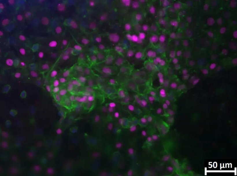
The Chang Laboratory's research focuses on the self-assembly of induced pluripotent stem cells (iPSC) in microgravity. These initial cells are created from normal human cells reprogrammed to act like embryonic stem cells. This means iPSCs can change into many different types of cells. These stem cells are built into liver tissues in microgravity that function like a smaller, simpler liver. Unlike Earth-bound tissue engineering methods that rely on exogenous matrices or culture plates, microgravity allows cells to float freely and organize naturally, resulting in more physiologically accurate tissues. A central component of this project is the development of a custom bioreactor, dubbed the "Tissue Orb," designed to facilitate tissue self-assembly in the weightless environment of space. The bioreactor features an artificial blood vessel and automated media exchange, simulating human tissues’ natural blood flow process.
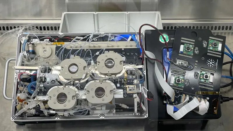
Image source: ACS; photo credit: Tammy T. Chang, MD, PhD
The research team is also working on advanced cryopreservation techniques to transport engineered tissues from space to Earth safely. The project’s next phase involves testing isochoric supercooling, a preservation method that maintains tissues below freezing without damaging them. This technology could extend the shelf life of engineered tissues and potentially be applied to whole organs. “Our goal is to develop robust preservation techniques that allow us to bring functional tissues back to Earth, where they can be used for a range of biomedical applications, including disease modeling, drug testing, and eventually, therapeutic implantation,” Dr. Chang said.
The Chang Laboratory's spaceflight experiment is scheduled for launch in February 2025. The authors maintain that the research not only showcases the potential of microgravity for advancing tissue engineering but also lays the foundation for future innovations in space-based biomedical manufacturing.
The research is supported by the National Science Foundation (NSF) in collaboration with the International Space Station National Laboratory (ISSNL) and the Translational Research Institute through NASA. Co-authors are Juan C. Reyna, MD; Anthony N. Consiglio, PhD; Alan Maida, BS; Maria Sekyi, PhD; and Boris Rubinsky, PhD.
Source: American College of Surgeons
21.10.2024