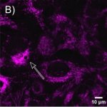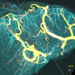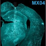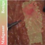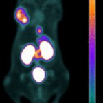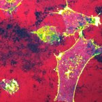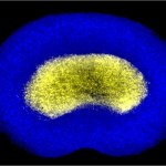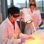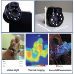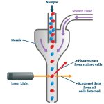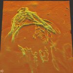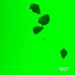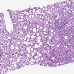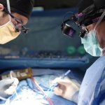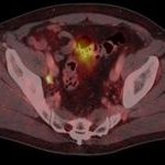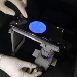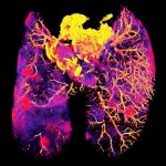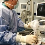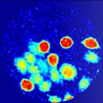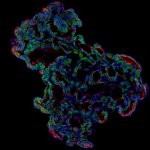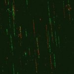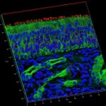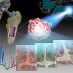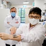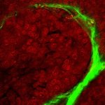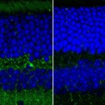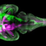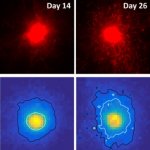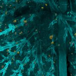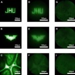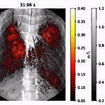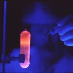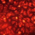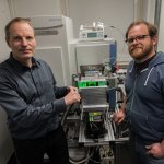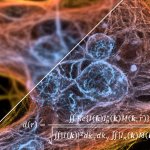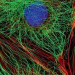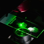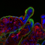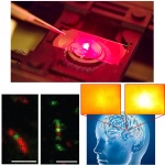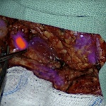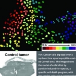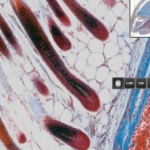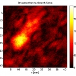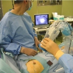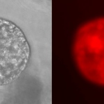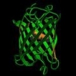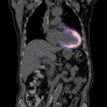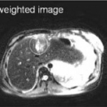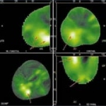
News • Fluorescence lifetime imaging microscopy
New technique could aid targeted lung cancer treatment
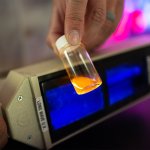
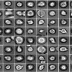
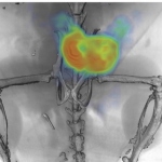
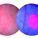
Detecting genetic mutations associated with lung cancer via laboratory tests can be expensive and time-consuming, and use up valuable tissue from small biopsy samples. A new approach aims to fix this.