Virtual slide for real analysis
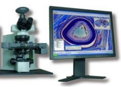
The updated Olympus dotSlide digital virtual microscopy system can scan entire slides at high resolution and fidelity, making them accessible and fully navigable anywhere in the world.
Using any of the three models - dotSlide MD (manual); dotSlide SL (fully automated, with slide loader), and dotSlide TMA (with a tissue micro-array module) - users can examine a virtual slide as if seeing the original through a microscope.
This allows pathologists and researchers, for example, to review cases without being near a microscope. Second opinions become quicker, and remote consultations, training and discussion possible, Olympus points out. The technology also provides high throughput and high content capabilities.
The dotSlide models use the Olympus BX51 microscope. Whilst with the dotSlide MD, slides and data are loaded manually and a virtual file created automatically, based on the users preferences, the automatic dotSlide SL has a slide loader holding up to 50 slides in 5 trays. A robotic arm places the slides on to the stage holder and the integrated barcode scanner ensures any bar-coded meta-data are automatically loaded and linked with the resultant virtual image, Olympus reports.
In research
The fluorescence compatible dotSlide can be used for tissue micro-arrays (TMAs) via the dotSlide TMA model. This facilitates the acquisition and simple analysis of tissue micro-arrays. TMAs consist of many small tissue cores with defined diameter that are fixed on a single slide. The model documents each core separately, accurately recording its slide and core reference for traceability.
For technology
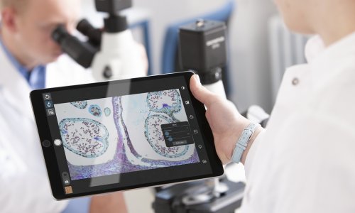
The dotSlide workstation and server system enable fully controllable remote access. Data and associated meta-data are saved in a bespoke data management system meaning that slides can be scanned in one location and reviewed almost instantaneously in another, anywhere in the world, by users either via a web browser.
Using the specialised OlyVia, software pathologists can review and comment on the virtual slide and are able to add annotations, markers and files (including dictations) to be directly linked to the slide. This software has many other assets and deserves to be followed up.
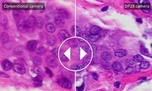
A Peltier cooled, 1376 x 1032 pixel dotSlide camera also offers high resolution, fast frame rates and very high sensitivity with an excellent signal-to-noise ratio, broad dynamic range and superior image quality.
31.08.2007