The Eisenherz project
Tobias Schaeffter PhD, Principal Scientist, Philips Research Laboratories, Hamburg, Arne Hengerer PhD, Director molecular MR, Siemens Medical Solutions, Erlangen, and Andreas Briel PhD, Eisenherz project manager, Schering Research Laboratories, Berlin, describe...

Recent developments in genomics and proteomics have tremendously increased our knowledge on the molecular processes of diseases. This feeds the hope that the susceptibility of an individual to many diseases can be predicted and that these diseases can be diagnosed at a much earlier stage and more individually designed therapies can be developed. Molecular imaging aims at the early detection and quantification of molecular processes associated with disease by using sensitive imaging methods together with targeted contrast agents. In order to make this vision happen, an interdisciplinary effort between different academia and industries is necessary.
The Eisenherz project is funded by the German Ministry of Education and Science (BMBF) to develop new targeted MR contrast agents and MRI technology that enables clinicians to detect cancer cells earlier, to stage the cancer, and ultimately, to quantify therapy effects of individual cancer treatments. This has massive implications for cancer management and could have sizeable cost-saving implications for hospitals.
In order to achieve these goals, Eisenherz has created an alliance for nanotechnology in cancer - a partnership between academic institutions, two of the leading medical imaging companies, Philips Medical Systems and Siemens Medical Solutions, and Schering, the leading contrast agent and pharmaceutical firm. The three-year research project, which began on 1 September 2005 under the leadership of Schering in the Nano-for-Life framework of the German Ministry of Federal Research, aims to translate German excellence in nanosciences to commercially viable medical products and processes.
To develop contrast-enhancing agents for molecular imaging is one major challenge facing the pharmaceutical industry today. Classic contrast agents primarily document the anatomy. For pathophysiological examinations using differential diagnostic techniques, in other words, characterising the development of a disease, they are only suitable to a limited degree. Molecular imaging selectively tracks down molecules and cell structures to be able to establish proof of diseases at a very early stage - and then to make decisions on a highly individual treatment.
The next straightforward vision of medical imaging quite clearly lies in the concept ‘Find, Fight and Follow’. In radiopharmaceuticals we are already pursuing the approach of a triad consisting of early diagnosis, therapy and therapy control. Building a bridge between therapy and diagnosis opens the field of ‘Theranostics’. With a ‘Find, Fight and Follow’ strategy, the tissue of interest first can be imaged via target-specific contrast particles. In a second step, the same particles, now combined with a pharmacologically active agent, can be used for therapy. And finally, monitoring of treatment effects is possible by sequential imaging.
Beside radiopharmaceuticals, utilising the nanotechnological concepts of colloid- and interface science imaging on a molecular level can also be achieved via MRI using tiny magnetic iron oxide particles coupled to target-specific ligands. Schering has many years experience in the field of contrast agent development and an already approved nanoscaled iron oxide MRI contrast agent called Resovist, in the market since 2001.
The device manufactures major strategic aims include supporting R&D in molecular medicine through the development of new ultra-sensitive imaging systems capable of observing phenomena at the molecular level, supporting the work of academic groups, and forming collaborations with contrast-agent and pharmaceutical companies.
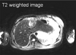
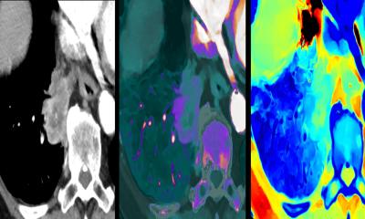
In particular, sensitive detection of a contrast agent in large volumes has to be developed to detect cancer cells with targeted contrast agents. In addition, the development of quantitative MRI is seen to be crucial for assessing disease progression or regression after therapy. Classical MRI usually allows only qualitative assessment of pathology by obtaining T1, T2 or T2* weighted images. By contrast, the mapping of relaxation times provides a quantitative estimation of the local contrast agent concentration. In addition, pharmacokinetic (PK) modelling is a valuable tool for objectively assessing contrast agent uptake in tissue. For this, mathematical schemes are developed that represent complex processes within the body. Quantitative MRI together with advanced PK modelling gives a valuable indication of, for example, the behaviour (uptake rate) of a tumour before and after therapy. Therefore, this technology has a high potential for an objective evidence of the effectiveness of therapy and can be used for individual dosage.
The project focuses on breast and prostate tumours - the most relevant cancer types. Therefore, targeted nanotechnology based contrast agents, common among these cancer types, will be developed by Schering in collaboration with the Max-Planck Institute of Colloids and Interfaces (Prof. Antonietti) and the start-up company Capsulution NanoScience AG. Regarding the dedicated tools and methods to characterise nanoscaled systems, the project will be supported by NanoLytics GmbH - a Berlin-based nanotechnology characterisation laboratory.
The developed contrast agent will be tested at three different hospitals for the different cancer types. The University Hospital Muenster (Professors Heindel and Bremer) will work mainly in the field of breast cancer. The detection of cancer cells will be tested and cross-validated with optical fluorescence tomography on animals. The Technical University (TU) Munich (Professors Schwaiger, Wester and Botnar) will focus on the detection of prostate cancer cells and will cross-validate the detection with positron emission tomography. The German Cancer Research Centre Heidelberg (DKFZ, Prof. Semmler, Dr. Bock) is evaluating the newly developed nanoscale contrast agents on various cancers models and will develop new animal MR-coils for clinical MRI scanners.
Since the hospital Muenster and TU-Munich are working with Philips equipment, these two sites are closely collaborating with Philips Research in Hamburg, whereas the DKFZ is collaborating with Siemens Medical Solutions in Erlangen. Both imaging industry partners (Philips Medical Systems and Siemens Medical Solutions) are working on standardisation issues, e.g. reference measurements for new, targeted contrast agents, and their limit of detection.
Eisenherz is a unique interdisciplinary consortium for the improved cancer management by means of Molecular Imaging: the early detection, the quantitative diagnosis and the selection of an appropriate treatment of cancer. This integrated approach has a high potential to improve the quality of life in future.
02.08.2006


