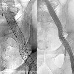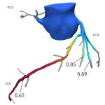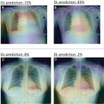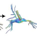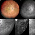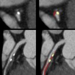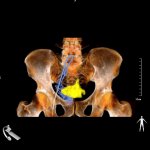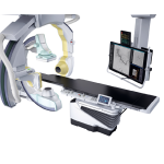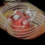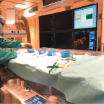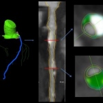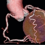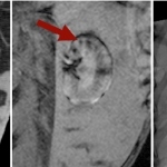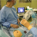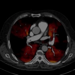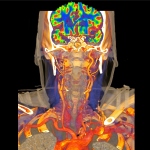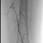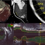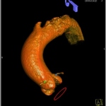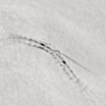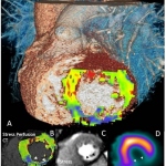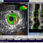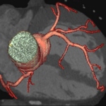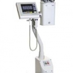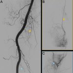
News • Genicular artery embolization
Lasting relief for knee pain – without surgery
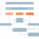
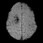
For patients with knee osteoarthritis, for whom injections no longer provide sufficient relief, but joint replacement is also not an option, a minimally invasive treatment might offer an alternative.