Breakthrough with new interventional treatment technology
Preliminary data from the clinical study showed that the dose reduction achieved in the X-ray guided endovascular procedures performed on 50 patients using Philips’ ClarityIQ technology is in line with the expected X-ray dose reduction of 75%.

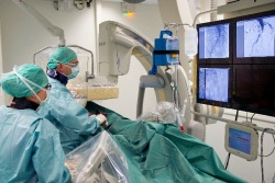
The St. Antonius Hospital Utrecht/Nieuwegein, the Netherlands, and Royal Philips Electronics (NYSE: PHG, AEX: PHIA) today announced an important breakthrough in the image-guided treatment of vascular patients. Using an innovative X-ray technology developed by Philips, the hospital’s interventional radiologists were able to dramatically reduce the amount of X-ray radiation that patients are exposed to during image-guided endovascular procedures without any loss in image quality. Preliminary data showed that the dose reduction is in line with the expected X-ray dose reduction of 75%. The clinical study involved a catheter treatment for narrowing of the iliac arteries due to atherosclerosis. The final results of the study, which was conducted on 50 patients, are expected in the first half of 2013, but the doctors at the St. Antonius Hospital are already considering the preliminary results an important medical breakthrough.
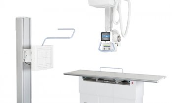
X-ray interventional systems are generally accepted as the most suitable imaging technology for accurately visualizing the progress of the catheter and other interventional tools during the minimally invasive treatment of cardio- and endovascular conditions. Philips is at the forefront of helping clinicians and staff to manage X-ray exposure to both their patients and themselves. The company is currently conducting worldwide clinical studies to study how its newClarityIQ technology can reduce the amount of X-ray radiation required to guide different kinds of minimally invasive treatments. The St. Antonius Hospital is the first hospital in the Netherlands to use this new type of X-ray interventional system.
“We did not have to fall back on the existing interventional system, which uses a normal dose of X-ray radiation, with any of the 50 patients. We could see that the images obtained with the new system were just as sharp. It enabled us to limit the X-ray dose administered to patients during treatment,” says Dr. Marco van Strijen, interventional radiologist and head researcher for the study at the St. Antonius Hospital Utrecht/Nieuwegein.
During this image-guided treatment, the interventional radiologist inserts a catheter into the relevant artery through a small opening in the groin and injects a contrast medium into it to make the blood vessels visible on the X-ray images. For a direct comparison between the new technology and conventional technology, the first series of images was captured using one of Philips’ conventional interventional X-ray systems and the second series using the new Philips AlluraClarity system with ClarityIQ technology, which sharply reduces the X-ray exposure required to produce the images. The experimental results were obtained by measuring the radiation dose received by the patient and by the radiologist, while wearing a lead apron throughout the procedure.
“All patients treated via X-ray guided interventions benefit from the advantage of reduced radiation exposure, but it is especially important when you are treating young adult patients and patients who have to undergo lengthy and complex procedures. Moreover, people with vascular problems often undergo the same type of treatment several times. With these reduced radiation levels, it is now in theory possible to treat a patient four times with the same amount of X-ray radiation that was previously required for a single procedure,” says Dr. Van Strijen.
Every year, some 1,200 patients receive endovascular treatment in the hospital’s radiology department. In the Netherlands alone, there are tens of thousands of patients who could benefit from this breakthrough.
Philips’ new X-ray technology is also being used for image-guided treatments in other parts of the human body - for example, for the treatment of blocked arteries in the brain (ischemic stroke) and pulmonary artery disease, or the image-guided cryo-ablation of tumors. The technique can also be applied during percutaneous coronary intervention (PCI) to treat blocked coronary arteries. A study was also completed recently at the Karolinska Hospital in Stockholm (Sweden). The results of this study show that during neurological treatments, the new Philips technology also provides similar image quality with a dose reduction of 73 percent.
“It used to be a technical challenge to combine high image quality with a low dose during X-ray guided interventions and this often gave rise to compromises,” says Bert van Meurs, General Manager of Interventional X-ray systems at Philips Healthcare. “We are proud that in close cooperation with our clinical partners we are working on a breakthrough that could make a substantial contribution to improving patient care. Live X-ray guidance is increasingly being used to perform minimally invasive treatments, which is why reducing X-ray dose without any loss in image quality is so important.”
18.12.2012
- angiography (117)
- imaging (1640)
- medical technology (1554)
- minimal invasive (201)
- non-invasive (82)
- radiation protection (187)
- X-ray (297)


