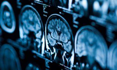
Polytrauma imaging
To be effective, guidelines, protocols and algorithms are essential
Professor Ulrich Linsenmaier, a leading expert in emergency radiology, has highlighted the need for clinicians to read image data rapidly in an emergency department if they are to help improve clinical outcomes for polytrauma patients.
Whilst he acknowledges that the volume image reading (VIR) approach advocated by his Munich group may be somewhat controversial, he believes that reading large, whole-body CT data directly on the console, prior to the arrival of reformatted images on the PACS, dramatically speeds up diagnosis.
Professor Linsenmaier, who heads the Institute of Diagnostic and Interventional Radiology at Munich Hospital KMPP and is an associate professor at Munich’s Ludwig-Maximilians-University, will present his views to the ECR 2013 Special Focus Session Imaging in Intensive Care Patients*.
Among other points, he will emphasise the importance of having clear guidelines, protocols and algorithms in place to ensure effective and safe imaging of polytrauma patients. He believes having such a structure available to the emergency department – along with the necessary imaging – will help speed up diagnosis, reduce complications and improve outcomes for the most severely injured patients.
Additionally, Professor Linsenmaier, who also presides over the European Society of Emergency Radiology (ESER), will focus specifically on polytrauma imaging, highlighting radiological diagnosis of polytrauma patients in his lecture covering logistics about radiologic management, scan protocols, imaging guidelines and patient care and follow-up imaging while being treated in intensive care units.
Speaking to European Hospital ahead of the conference, he said there are a number of key factors to consider to successfully set up an emergency radiology department to treat polytrauma patients. The first step, he explained, is triaging patients for the emergency department treatment (ED), following Advanced Trauma Life Support (ATLS)-based entry criteria to securely establish whether the patient has polytrauma. ‘Next, you need clear algorithms for early clinical treatment in the emergency room with radiology being part of the ED team. Good communication between radiology, anaesthesiology, emergency medicine and surgery is essential.’
A third step is to use ultrasound for Focused Abdominal Sonography for Trauma (FAST). Professor Linsenmaier and his team having defined a modified FAST version, which they prefer to use – it also quickly views the heart and bilateral pleural spaces for haemorrhage. FAST is a critical step in how quickly a patient can undergo CT, and whether they are triaged directly from the emergency room to the operating theatre.
He stressed that the radiologist on-site in the emergency room must have specific emergency radiology knowledge; the technologist running the CT scanner should be familiar with performing whole body scans in polytrauma; and there needs to be a pre-programme protocol, specifically designated to whole body CT.
Of his firm belief in the VIR concept having particular value in this context, he further points out: ‘Today’s whole body CT produces a large number of images, with which it’s difficult to work, so my group recommends VIR - volume image reading – where we start reading the data before the images arrive in the PACS. This is new, even a little controversial, but an interesting concept that speeds up radiology because we can read the images right away when the data is available on the CT console. We don’t wait until every reformatted image is in the PACs, we instead start VI reading the raw data immediately from the console and this speeds up the diagnosis.’
With a limited role for conventional radiography in this context, the professor recommends having the availability of ‘a modern, very fast scanner, capable of handling large scan volumes and ensuring integration of all modalities’ for successful polytrauma imaging.
The key learning points from the ECR session, he said, will be how to apply modern emergency radiology in a clinical priority-oriented concept. Adopting such processes will speed up diagnosis, he added, reduce the complication rate and improve the outcome for the patient.
The professor’s Munich team has conducted research on the influence of whole body CT in polytrauma on patient outcomes. ‘We showed that applying whole body CT in patients with polytrauma, significantly improves survival,’ Professor Linsenmaier confirmed (The Lancet. 2008).
As for the latest developments, he believes many advances have already been made with today’s scanners in assessing vascular and head and neck injuries with the option, for example, to perform a CT angiography of the supra-aortic arteries to rule out vascular injury.
The ECR session will also examine point-of-care versus diagnostic ultrasound in the intensive care unit.
Professor Linsenmaier is keen that advances in polytrauma imaging are not only available in major centres but that the knowledge, teaching and training cascades down to rural areas to improve diagnosis in polytrauma in less specialised radiology departments.
* Friday, 8 March. 16:00-17:30. SF 7c: Imaging in intensive care patients.
PROFILE
Professor Ulrich Linsenmaier is Head of the Institute of Diagnostic and Interventional Radiology in Munich Hospital and Associate Professor at Ludwig-Maximilians University, Munich. He is also President of the European Society of Emergency Radiology (ESER) and has carried out extensive research on the benefits of CT scans for seriously injured patients as well as techniques for imaging polytrauma cases.
27.02.2013