CT will remain an imaging heavyweight
Computed tomography (CT) is the modality of choice for many diagnostic issues. Whilst currently its major strength is the visualisation of anatomical detail, future technological improvements may also reduce radiation exposure.

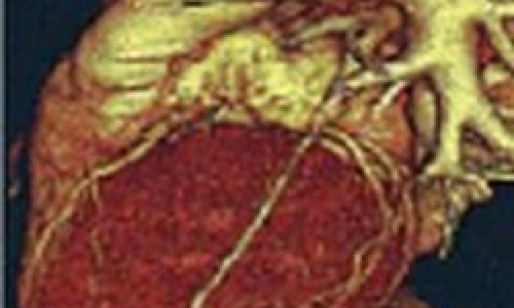
‘Today, computed tomography is the best imaging modality to detect stenoses in patients with intermediary pre-test probability where a coronary angiography is not immediately indicated,’ according to Professor Dr Gabriele A Krombach, Department Director at the Clinic of Diagnostic and Interventional Radiology at University Hospital Giessen and Marburg (UKGM). The degree of anatomical detail, she adds, is much better in CT than in magnetic resonance imaging (MRI). ‘In malignant variations of the descending artery, where the coronary artery is compressed between aorta and pulmonary artery, CT is currently the diagnostic method of choice.’
CT is also the modality of choice for visualising coronary stenoses. With low-risk patients without suspected coronary stenoses who require cardiac surgery, e.g. to remove a cardiac tumour, a CT scan can provide valuable information on possible coronary stenoses and help predict surgical outcome.
A further important indication for CT is the visualisation of plaque to risk-stratify patients with medium pre-test probability, Dr Krombach adds. ‘With an asymptomatic patient who has a risk of 10 to 20 percent to develop coronary heart disease over the next ten years, quantification of coronary calcification with CT in indicated.’ However, with higher risks CT is not indicated and the patient has to undergo an invasive angiography right away. Similarly very low risk (below 10 percent) asymptomatic patients do not need a CT. ‘In such a cases,’ she explains, ‘we simply wait and see.’
The radiologist particularly appreciates CT in percutaneous aortic valve replacements (TAVI), recently experiencing a veritable boom. According to the Herzbericht 2010 by Dr Ernst Bruckenberger, the number TAVI procedures increased from 93 in 2006 to more than 4,800 in 2010. ‘When planning a percutaneous aortic valve replacement the diameter of the ring and the distance of the coronary artery ostia to the valve have to be determined – this is done best with CT,’ Dr Krombach explains. As a radiologist she does not expect CT to fall into obscurity any time soon, primarily because no single modality can do everything. The coronaries, for example, cannot be visualised in echocardiography. Additionally, technological progress in CT might open access to new patient groups. The alpha and omega of CT development is the reduction of radiation exposure and indeed new kinds of detectors are being designed that aim to decrease radiation to submillisievert level.
‘If we could realise dose reduction in CT by using the different spectra of X-rays, we could also examine younger patients, such as those with congenital heart defects’ – patients who today undergo MRI, she stresses.
Frequent indications for CT
• Patients for cardiac surgery not involving the coronary arteries, such as valve replacement or cardiac tumour resection
• Patients with intermediate risk who should not undergo a coronary angiography
• (Suspected) coronary anomalies
• Evaluation of bypasses (problems: calcification of the native vessels, evaluation of the anastomoses), also in case of repeat surgery to visualise existing bypasses
• Method of choice for percutaneous valve replacement
• Visualisation of cardiac veins prior to the implementation of a bi-ventricular pacemaker
• Visualisation of the pulmonary veins prior to ablation with arrhythmias
• Visualisation of the pulmonary veins post ablation (suspected stenosis)
• Anomalous pulmonary venous connection
Profile:
Professor Gabriele A Krombach MD studied medicine and specialised in radiology at Aachen medical school, later receiving her teaching certification for diagnostic radiology in 2003. In 2006 she became chief senior resident at the Radiological Diagnostics Clinic at Aachen University Hospital and was appointed adjunct professor in 2008. Two years later she became Director of the Diagnostic and Interventional Radiology Clinic at University Hospital Giessen, where cardiac imaging, MRI, interventional radiology and pulmonary imaging are her research priorities.
26.08.2013