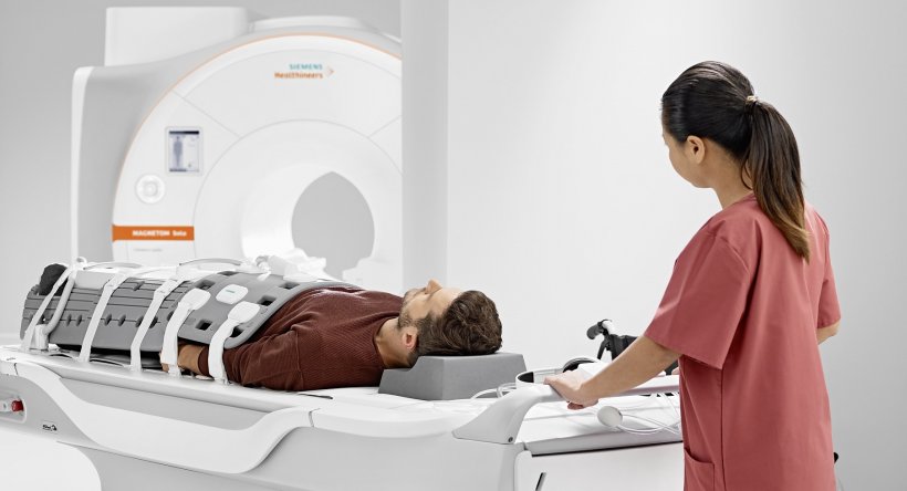
Source: Siemens Healthineers
Article • Innovation
AI helpers simplify clinical MRI scans
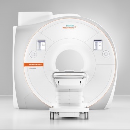
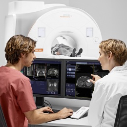
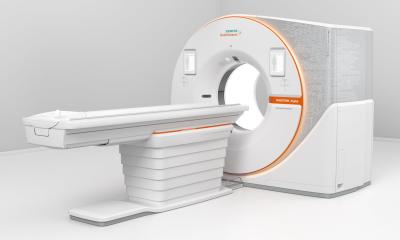
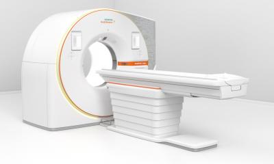
The new 1.5 Tesla MRI from Siemens Healthineers, Magnetom Sola, is packed with helpful algorithms and other functions. AI-supported systems monitor patients and scan parameters and ensure consistent image quality. Whilst visitors at this year’s ECR-Expo admired the new device, Prof. Ulrike Attenberger has already tested it in practice.
Report: Wolfgang Behrends
The Vice Chair at the Institute of Clinical Radiology and Nuclear Medicine, University Medical Centre Mannheim, talks about the effect of the new functions in clinical routine.

With long-term investigations in particular, this reduces the comparability of data enormously, even for the same patient in the MRI scanner. Different patients’ individual characteristics also cause additional inconsistencies: ‘Obesity impacts on image quality and so, does arrhythmia, for example. Some patients cannot hold their breath, which used to cause us huge problems.’ The new technology also helps the examiners here. ‘We enter patient data into the system, i.e. height and weight, how long they can hold their breath, and then the algorithm calculates the settings that will achieve the best image quality,’ Attenberger said. ‘This has worked very well during the first practice runs.’ The manufacturer already implemented the new functions in the 3-Tesla System Magnetom Vida. ‘However, the introduction in the 1.5-T range makes the technology available to a much larger range of users, especially in clinical practice,’ added Dr Christoph Zindel, Senior Vice President and General Manager of MRI at Siemens Healthineers.
Even MRI novices adapt quickly to the system
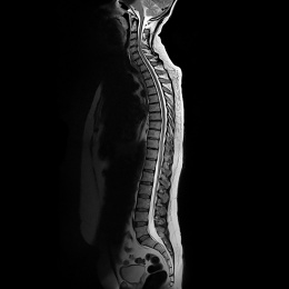
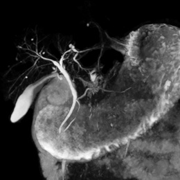
‘During the test phase,’ Attenberger pointed out, ‘we made a point of selecting our most junior MRI assistants who are being coached by skilled assistants but don’t yet have much MRI experience. After three days of training on the simulator they then used the device in practice, and it turned out the handling is so intuitive that there were no problems during the introduction.’ The first test phase began with only one of the three sensors, i.e. the respiratory sensor, and even just this one component convinced Attenberger and her team. ‘The system works perfectly in practice. Movement is always a big problem for us: patients in our centre are becoming increasingly older and sicker, which leads to high fault rates through motion artefacts with conventional MRI scanners. The BioMatrix technology now counters this problem very effectively. The new system makes the image quality far better.’
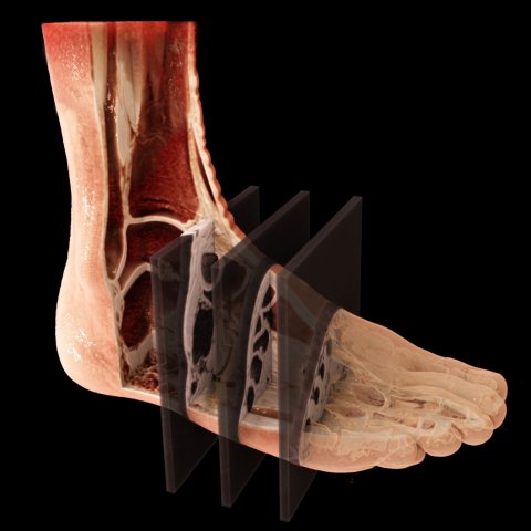
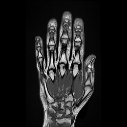
It also has a significant impact on the examination time: ‘We aim to reduce the time spent in the examination room by around 50%, from an average 50 minutes to 25 minutes. Having used the new device for the first few weeks now, we can foresee this should be achievable.’ The new simultaneous multi-slice technology also contributes towards this because it facilitates simultaneous images of several slices. This function really excels during musculoskeletal examinations. In cardiac and abdominal imaging, patients benefit from compressed sensing. The technology enables the examination of cardiac function with free breathing through mathematical reduction of the acquired data. ‘You simply start scanning and continuously acquire images over a three-minute period, without the need for the usual breath-holding commands,’ Zindel observed. ‘Afterwards, the intelligent algorithm automatically selects the best images from this data jungle.’
Wish list during development
It is no coincidence that the Mannheim-based radiologist is currently carrying out the test run with the Magnetom Sola. Her institute collaborated with Siemens Healthineers during the conception of the new device and provided input for the developers. ‘We contributed our ideas and requirements from a clinical routine perspective,’ Attenberger said. ‘This type of cooperation with Siemens Healthineers has a long tradition.’ Is she pleased with the result? Yes. ‘All our suggestions have been implemented.’ Along with the new functions these also include connection to the Teamplay software, which delivers an analysis of the clinical processes and optimises MRI capacity.
Profile:
Professor Ulrike Attenberger is Vice Chair and Medical Director at the Institute of Clinical Radiology and Nuclear Medicine, University Medical Centre Mannheim. After gaining her medical degree in Munich, a doctorate focused on ‘The importance of MRI in the diagnosis of pulmonary hypertension’ followed in 2006. Her current research is on MRI for tumour diagnosis and capturing therapy response. The professor received an RSNA Fellow Award for her work on optimisation and dose reduction for contrast-enhanced MR angiography.
11.11.2018


