Article • Teamwork <3
7-Tesla: Multidisciplinary care is key to cardiac disease management
New 7-Tesla MR methods could potentially shed light on cardiomyopathies’ principles, according to a leading French radiologist who also stresses the importance of teamwork between radiologists, cardiologists, surgeons and anaesthesiologists.
Report: Mélisande Rouger
Morphologic and dynamic information of the myocardium is achieved with millimetric resolution (0.9x0.9 mm2). Strong intensity variations characteristic of 7-Tesla MRI can be observed from anterior to posterior myocardial segments.
New tools provided by industrial partners and used by cardiovascular surgeons and radiologists are improving treatment of thoracic aorta pathologies. An increasingly used technique is fusion imaging, in which pre-treatment MR and CT scans of the patient are being fused with angiography images to guide stent-graft navigation through the vascular structures of the patient during the intervention, according to Alexis Jacquier, cardiovascular radiologist at Timone University Hospital in Marseille. ‘Fusion imaging enables to lower radiation dose and to reduce the amount of contrast media that are traditionally required in this type of surgery. It avoids having to inject iodine to know where we’re at,’ he explained.
The hospital also hosts the Timone Aortic Centre (CAT in French), a leading regional multidisciplinary centre that covers full aortic pathology management, from diagnosis to patient care and follow-up, with a strong connection with the university. The CAT includes vascular and cardiac surgeons, radiologists, cardiologists, vascular physicians and anaesthesiologists. The objective is to provide a multidisciplinary approach to provide the best medical care; for instance all thoracic stent-graft procedures are performed by a multidisciplinary team comprised of vascular surgeons, radiologists and anaesthesiologists at CAT.
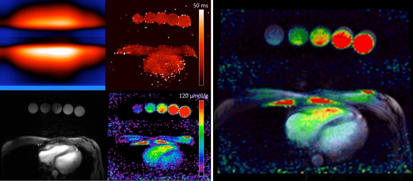
Figure 1 (left): Top-left is B1+/B1- map, Top-right is T2star map, Bottom-left is anatomical MRI image of the heart using conventional 1H-MRI, and bottom-left is the map of long-relaxation sodium concentration after corrections of the sodium MRI signal using the maps at the top.
Figure 2 (right): Overlay of long-relaxation sodium map onto the anatomical MRI.
Imaging has become key in thoracic aorta treatment with the boom of minimally invasive procedures. Besides thoracic disease, Timone Hospital is one of the main centres in France offering endovascular interventional radiology skills to treat patients with carotid and renal disease, which Jacquier and colleague Vincent Vidal perform daily, along with the full suite of cardiovascular interventional radiology procedures – endoprostheses and stent placement, small vessels and tumour embolisation, etc. Furthermore, the hospital is located close to the medical and biology MR centre (CRMBM), one of the few labs in Europe that work with 7-T MRI for diagnostic imaging research.
This proximity enables Jacquier and team to test 7-T methods using sodium instead of proton imaging, a possibility that opens brand new perspectives in heart imaging. ‘Sodium electrolytic disorganisation in the myocardium can have an electrical and mechanical impact on heart function. 7-T will enable the development of new applications in the field. It is still a complex task, but we are working hard on different papers on sodium quantification in the myocardium and potential clinical applications’ he explained.
Cooperation with cardiologists is essential in myocardial disease management, according to Jacquier, who again stressed the importance of the multidisciplinary approach during patient treatment. ‘Patients are now being care for within the heart team, a model increasingly followed by healthcare facilities in France and beyond,’ he explained, adding, ‘whether it’s for TAVI procedures, diagnosis or follow-up. Medicine is becoming hyper specialised and mixing profiles and specialties enables us to significantly improve patient care.’
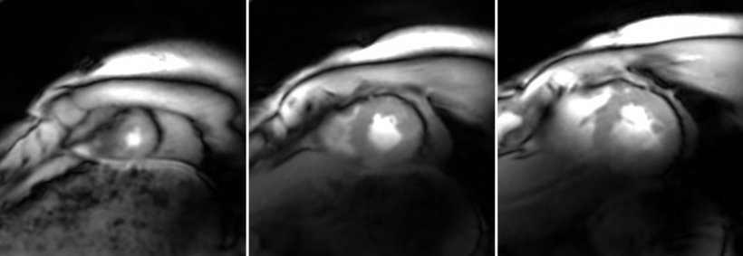
Figure 3: SMS cine acquired within 10s showing diastole (relaxed phase of the cardiac cycle) of the apex (a) mid-ventricular (b) and base (c) slices.
Another significant development in France was the reform of the radiology residents’ training scheme, which was introduced in 2017. Radiology residents must now undergo a three-step training, including successively: base training (one year), dedicated to emergency radiology; in-depth training (three years), to ensure that every subspecialty in radiology has been covered in their education; and consolidation training (one or two years), providing certification for one or two subspecialties.
The French Council of the Teachers of Radiology (CERF) has been piloting the change for radiology. The French Society of Cardiovascular Radiology now provides e-learning material to ensure homogeneous teaching and training program across the country. In September, the series became freely available for French residents on the CERF website, and also available for all radiologists on the website of the French Society of Radiology.
This change is a substantial improvement in the training scheme, because it reflects daily routine better, Jacquier added. ‘Cardiac imaging studies are being prescribed every day by all sorts of physicians: GPs, endocrinologists, surgeons, and even oncologists, for instance in pre- and post-chemotherapy evaluation.’
A lot of things may need to be updated as we gradually introduce artificial intelligence
Alexis Jacquier
As for cardiology, the French Society of Cardiology and the French Society of Radiology established a working protocol in 2005; according to this, the cardiologist prescribes the CT and MR scans and radiologist performs the technical assessment and writes the report – and then sends it to the cardiologist. Jacquier: ‘This division of tasks promotes the best possible medical care, but everything really depends on the physician’s skills. A lot of things may need to be updated as we gradually introduce artificial intelligence.’ Radiologists must also homogenise the way they write the imaging report. Introducing the structured report to exploit data at national level will prove essential for their future. Another priority is to improve communication not only with patients but also other medical specialties, he said.
Jacquier will participate in the International Day of Radiology (8 November 2018), an initiative to highlight the radiologist’s role in cardiac care. ‘Radiology is not a medico-technical specialty, although French administration still classifies us as such. We’re a medical discipline. The old-fashioned image of the radiologist reading scans alone in a basement and not having contact with anyone else in a hospital is outdated. The radiologist,’ he emphasised, ‘is now at the centre of patient care and healthcare.’
Profile:
Cardiovascular radiologist Professor Alexis Jacquier, at Timone University Hospital, Marseille, France, trained in Marseille and Lyon and gained his PhD in San Francisco, USA, supervised by Maythem Saeed and Charles Higgins. In 2006 he integrated the cardiovascular group in the CEMEREM research lab (http://crmbm.univ-amu.fr). He is author and co-author of more than 90 peer-reviewed publications and has presented numerous lectures, tutorials and refresher courses internationally. He also chaired the European Society of Cardiac Radiology membership committee and is current vice president of the French Society of Cardio-Vascular Radiology (Société Française d’Imagerie CardioVasculaire, SFICV).
26.11.2018