News • Blindness prevention
Blue is the clue for diabetic retinopathy risk
Researchers from Tokyo Medical and Dental University (TMDU) demonstrate a thorough and non-invasive imaging technique to identify areas of the eye affected by diabetic retinopathy (DR), a progressive eye disease associated with diabetes and a leading cause of blindness.
Image source: Unsplash/Tengyart
The researchers have found that blue light can be used to probe the depths of the eye and uncover areas affected by DR. In a new study published in Asia Pacific Journal of Ophthalmology, they have revealed that blue images obtained by multicolor widefield scanning laser ophthalmoscopy (SLO) may be used to identify areas of DR-induced damage in a more extensive portion of the eye compared with previous methods.
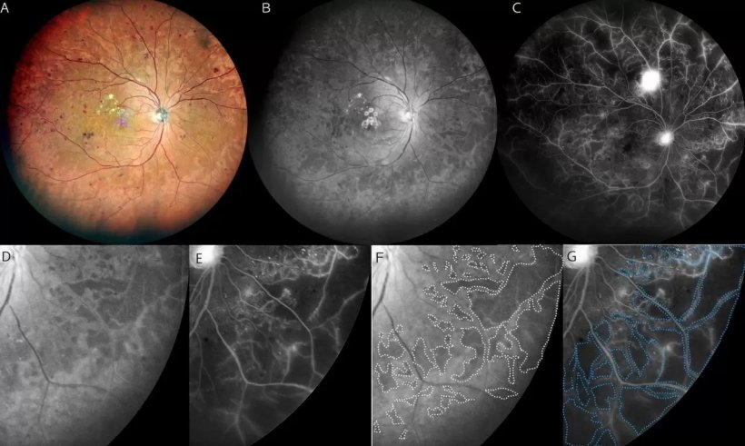
A: Multicolor widefield SLO image of the right fundus of a 65-year-old man with PDR showing multiple hemorrhages in a wide area of the fundus. B: Blue SLO image shows a hyporeflective area in the mid-periphery to periphery of the fundus. C: Widefield FA image shows widespread NPAs in the mid-periphery to periphery. Neovascularization is also seen in the superior pole of the eye. D: Magnified image of image B shows hyporeflective areas in the lower temporal quadrant. E: Magnified FA image of image C shows NPAs in the same quadrant of image D. F: The hyporeflective areas in image D are outlined by white dots. G: The NPAs in image E are outlined by blue dots. The outline of white dots in image F is located inside the outline of blue dots image G.
Image source: Horie et al., Asia Pacific Journal of Ophthalmology 2021 (CC BY-NC-ND 4.0)
Current eye imaging methods include fluorescein angiogram, which involves the injection of dye into the eye. SLO is a non-invasive approach that does not require dye, and multicolor widefield SLO represents an advancement of this technique in which red, blue, and green lasers are used to simultaneously capture images of a wide portion of the eye. Previous research has shown that blue images captured by conventional SLO may reveal hyporeflective areas in the eye indicative of damage associated with DR. Researchers at TMDU sought to further evaluate this finding using widefield SLO.
In this retrospective study, the researchers compared blue widefield SLO images and fluorescein angiogram images taken in people with diabetes. The morphology of the retina was also evaluated in some individuals with DR. “We found that the hyporeflective areas in the blue widefield SLO images appeared to correspond with areas of ischemia in the fluorescein angiogram images of patients with DR,” explains Kyoko Ohno-Matsui, senior author. “We were pleased to find that the rate of concordance was high.”
Further evaluation of patient images showed that ischemic areas (i.e., areas of reduced blood flow) appeared to correspond with parts of the retina that were thin and partially disorganized. “It’s possible that the blue wavelength of light can pass more easily through these thinned areas of the retina, which presents as hyporeflective areas in the SLO images,” says Horie.
This study confirms the utility of blue widefield SLO as a simple and non-invasive tool for the detection of DR-associated damage in the eye. This technique may serve as an important means of screening and monitoring disease progression in individuals with DR.
Source: Tokyo Medical and Dental University
29.10.2021