Emerging and merging disciplines
Medicine as a profession has held a superior aloofness for many centuries, wary of losing its unique distinctiveness and esteem if ‘tainted’ with other professions.

However, in recent decades the pressure of progress and whirlwind of change has swept the Hippocratic race off its professional Olympus.
The affiliation of the medical profession with that of engineering has brought a new dimension in the diagnostic power of the clinician in many fields. The two-dimensional limitation of the X-ray has exploded in the 3-D reality of MRI and 4-D ultrasonography, while open surgery and exploration has been replaced by the less invasive and thus safer endoscopic procedures.
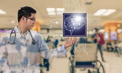
In the latter field, biomedical engineering - now a new branch in both professions - has moved a step further with the introduction of capsule endoscopy, where the patient is relieved of a highly uncomfortable procedure, instead swallowing a capsule containing a minuscule camera that can capture up to 35 images per second. The capsule communicates wirelessly with a receiver, which the patient comfortably carries at home for about eight hours, after which the capsule is passed out and the clinician assesses the images.
The clinician can use many methods to process and classify the images – manually, which is obviously very laborious and time consuming, or using one of the many systems of artificial intelligence to facilitate the process. Professor of Engineering Kenneth Camilleri and engineer Carl Azzopardi, respectively the director and biomedical engineer at the Centre for Biomedical Cybernetics, University of Malta, recently launched software whereby the processing and classification of endoscopic images can be facilitated. ‘The software can help the clinician in differentiating the mucosa of one gastrointestinal organ from another, thus classifying them and marking the valves (pylorus and ileocaecal) that are used for orientation,’ Carl Azzopardi explains. ‘With further adaptation, the software could eventually be made to go through the images automatically and pick up polyps, ulcers and tumours, if the right image features are detected and classified.’
This software is still in the research stages and, although preliminary studies look promising, there are still a number of teething issues with which to contend. ‘One of the questions is how much the clinicians will trust the software and how much they will take responsibility for diagnosis,’ Professor Camilleri points out. ‘Ideally, the software should be used as a diagnostic support and not replace the clinician’s acumen.’
This is not the only project on which the Maltese team is embarking. Another research programme aims to process ultrasound images from a regular 2-D ultrasound probe, in order to collate them together and create three-dimensional representations of vascular anatomy in peripheral sites, such as the carotid or femoral artery. Currently, whilst 2-D ultrasound modalities are well established in clinical practice they are less effective than their counterparts, e.g. CT or MRI angiography, especially when gauging low to moderate degrees of occlusions, where diffuse disease is present or where the patient suffers from cardiac arrhythmias,’ the two engineers explain. ‘Present 2-D ultrasound modalities are also not ideal for follow up studies, as 2-D imaging makes relocation difficult and is operator dependent.’
On the other hand, the team add that 3-D ultrasound probes themselves are very expensive and can only image a limited volume at a time, whereas present 3-D freehand techniques are qualitative at best, and have many artefacts.
The project therefore aims to develop a hybrid combination of in-built position sensors and image processing techniques, to use normal, cheap 2-D probes to generate accurate and ‘panoramic’ 3-D models of vascular anatomy, enabling anatomical spatial relationships to be observed. The next step would then be to study novel means of quantifying the volume of atherosclerotic plaques, and to compare the diagnostic confidence of such a method with gold standards such as CT or MR angiography.
Capsule endoscopy and ultrasound imaging are both fields within biomedical imaging that are receiving much attention. In fact, there are similar research programmes at EU other universities that probe into different aspects of these topics.
At the University of Porto in Portugal, research is taking place on using structured learning methods for assessing Barrett’s oesophagus using narrow band imaging in regular endoscopic techniques.
At the University Autonoma de Barcelona, researchers are using automated image processing techniques as a means to assess the intestinal motility of a patient using capsule endoscopy.
Meanwhile, at University College London, ultrasound imagery of the prostate is fused with 3-D MRI models to assist with pre-operative planning, whereas Cardiff University researchers are using 3-D Doppler ultrasound techniques to study neovascularisation in tendinopathy.
Shakespeare once asserted that, ‘there is nothing so becomes a man as modest stillness and humility’. The ‘sharing’ of the interpretative and investigative acumen of the medical profession with that of engineering can be well seen as a catalyst in that sometimes hazy world of diagnosis where one deviation can spell nothing less than a human life.
Profile:
Carl Azzopardi is a biomedical engineer at the Centre for Biomedical Cybernetics, specialising in clinical engineering, with a particular focus on medical imaging. He also heads the Biomedical Engineering Department at Saint James Hospital Group.
Professor Kenneth P Camilleri directs the Centre for Biomedical Cybernetics and works extensively on the application of signal and image processing to biomedical engineering, with a particular interest in brain signal analysis and brain-computer interfacing
16.05.2014