Dose discussion: How low can you go?
How low? During the GE Healthcare Lunch Symposium of GE Healthcare at this year’s ECR in Vienna, Michael Maher, Professor of Radiology at the University College, Cork, provided an answer: 1.2 millisievert – at least for abdominal CT scans in patients with Crohn’s disease.
“Crohn’s disease patients represent a group of patients that can need regular CT follow up exams and hence, lowering radiation dose associated with CT is vitally important in these patients. The question we studied at Cork University Hospital was how radiologists can use emerging CT technology to help these patients by significantly lowering the radiation dose per CT examination.
Other centres (e.g. Massachusetts General Hospital, Boston, MA, USA) have recently reported significant reductions in radiation dose in Crohn’s disease patients for CT scanning of abdomen and pelvis (CT ABD/PELVIS) using recently available Adaptive Statistical Iterative Reconstruction (ASiR). The MGH group reported a reduction in radiation exposure from an average dose of 7.7 millisieverts, to 5.6 millisieverts using ASiR. Our ambition was to investigate the feasibility of reducing the radiation dose of CT ABD/PELVIS much more significantly, to the level approaching that of a plain radiograph of the abdomen, which is approximately 0.7 millisievert. Again, to attempt to achieve that ambitious aim, like the MGH group, we also used ASiR, which is iterative reconstruction software that reduces noise, which is inevitably found on ultra-low dose CT images.
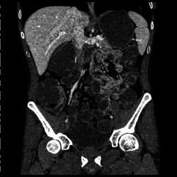
We found that ASiR successfully reduces the noise and therefore significantly improves the diagnostic acceptability of the ultra-low dose CT ABD/ PELVIS images. With the iterative reconstruction technology we were able to reduce the dose to a mean of 1.2 millisieverts, which we consider to be much more acceptable level of exposure in this patient group. Our study includes approximately 50 patients and was prospective, and all patients recruited had two CT scans: one at a mean effective dose of 4.7 millisieverts and a second ultralow dose CT ABD/PELVIS scan with a mean of 1.2 millisieverts.
This study design facilitated a comparison of a low dose scan with an ultra-low dose scan for image quality, diagnostic acceptability and ability to detect pathological findings. We found that ASiR satisfactorily reduced image noise and produced diagnostic images of reasonable quality. All pathological findings seen on the low-dose images were also detected on the ultra-low dose images. In our diagnostic imaging department, which serves a large tertiary referral centre, we use ASiR routinely for all CT scans, including CT scans of head, thorax, abdomen and pelvis and for peripheral and neuro CT angiography, and have been impressed by our ability to achieve radiation dose reductions while maintaining image quality.”
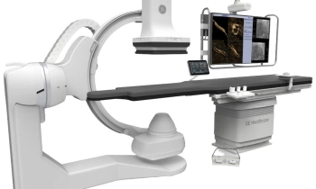
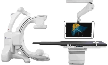
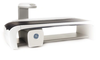
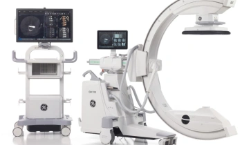
Adaptive Statistical Iterative Reconstruction (ASiR) by GE Healthcare
Typically, lowering dose has increased noise and image artifacts, creating an unfortunate trade-off between the high image quality and the low dose. To overcome this obstacle, GE has developed an industry breakthrough approach to image reconstruction — Adaptive Statistical Iterative Reconstruction (ASiR). ASiR is designed to enhance image quality, increase low contrast detectability (LCD) and remove noise without degrading anatomical integrity. It overcomes the limitations of the conventional CT reconstruction approach known as filtered back projection and arrives at an optimised image using an advanced iterative computation, directly from raw data. More than 900 GE sites are already using ASiR and more than 7,000,000 examinations have been performed using this technology.
20.06.2011
- CT (617)
- imaging (1676)
- medical technology (1580)
- monitoring (383)
- radiation protection (187)
- security (460)
- therapy (862)
- thorax (78)