Strategies for radiation dose reduction in vascular CT
Interview with Christian Loewe, MD, Professor in Radiology, Medical University Vienna, Austria
Radiation dose reduction in CT angiography can be achieved by reducing the kV settings, reducing the tube voltage, the tube current and by using iterative reconstruction algorithms.

Why we need radiation reduction in vascular CT?
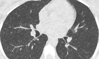
The exciting technical innovations and improvements in CT over the last decade have led to CT angiography becoming the method of choice for nearly all anatomical regions and clinical indications. CT angiography is nowadays the first choice technique in cases of ischemic stroke [1], for all aortic diseases (including acute aortic syndrome and treatment follow up) [2], for pulmonary embolism and for the diagnosis of abdominal vascular disease (e.g.mesenteric ischemia, and renal artery stenosis) [3].
Even in the peripheral arteries, an increasing number of studies have demonstrated the high clinical usefulness of this method for the management of patients with peripheral arterial occlusive disease [4-5]. Unfortunately, while the advantages of CT angiography are obvious in terms of patient management and disease diagnosis, an unavoidable drawback is the radiation dose required. This is particularly problematic for patients with chronic progressive diseases such as peripheral arterial occlusive disease (pAOD) which requires repeat follow-up imaging; in such cases the cumulative radiation dose is an important issue. In looking to address this issue several recently published papers have focused on the risk for radiation induced cancer in cardiovascular applications [6-7].
Why we can reduce radiation dose in vascular CT?
In general, there is always a compromise in CT between image quality (mainly defined by the image noise) and the radiation dose; the phrase: ‘more dose means “better” images’ was established at the beginning of the CT era and is still valid in some regards. However, in CT angiography, image quality is mainly determined by the contrast between the contrast-enhanced arterial lumen and the non-enhanced surrounding tissue.
Usually, this contrast is very high if contrast administration is performed in an appropriate way (see below). Thus, increased image noise is not really a problem in vascular CT, since the contrast will remain sufficiently high. Additionally, by reducing the kV settings – a powerful strategy for radiation dose reduction – the attenuation of iodine is increased leading to still greater contrast and thus a further decreased importance of the image noise due to the lowered kV [8]. Thus, radiation dose saving strategies are particularly appropriate in CT angiography. However, it is extremely important to optimize and adapt the contrast administration to these altered settings.
How can we reduce radiation dose in vascular CT?
The possibility to reduce radiation dose signif icantly by lowering the kV setting as discussed above is already established in many different anatomical regions. It has been shown to allow for a dose reduction of up to 50% [9-12]. The increased image noise due to the low kV settings (between 80 and 100 kV) was not considered a problem in any of the published studies due to the increased intraarterial signal intensity. Thus, despite the increased background noise, contrast-to-noise ratio was higher in the low dose protocols as compared to the standard (high) dose protocols as compared to the standard (high) dose protocols. However, lowering the tube voltage is only one of the currently available dose saving strategies.
Another approach is to also reduce the tube current (mAs), since there is a direct relationship between tube current and radiation dose. Although this approach will also lead to reduced image quality due to increased image mottle, this can be compensated for by new algorithms for image reconstruction: reduction of image nose by using iterative reconstruction techniques (instead of the “normal” filtered back-projection techniques) has been shown to allow the radiation dose to be reduced without sacrificing diagnostic confidence [13-14]. Finally the availability of high-pitch scanning techniques has demonstrated an additional dose saving potential in some dedicated anatomical regions and clinical applications, especially children and aortic imaging. If a high pitch technique is used then ECG triggering is no longer mandatory. This leads to further radiation dose reduction in aortic imaging [15-16].
How can we use these techniques in clinical imaging protocols?
The importance of appropriate contrast administration remains fundamental for the quality of CT angiography even in times of ultrafast and low-dose scanning. Due to above mentioned increase of image noise due to different dose saving strategies, intravascular contrast remains crucial to keep the contrast-tonoise ratio high enough for diagnostic image quality. Although there is as yet no clear definition about the level of CNR really needed for diagnostic image quality, the usefulness of using contrast agents that contain a high concentration of iodine has been confirmed in a number of publications [10, 17].
A combination of all currently available dose saving techniques (lowkV, lowmAs, Iterative reconstruction, high pitch scanning) with perfectly optimized administration of highly concentrated contrast agents allows for high-quality CT angiography at significantly reduced radiation dose.
####
References:
1. Latchaw RE, Alberts MJ, Lev MH, Connors JJ, Harbaugh RE, Higashida RT, Hobson R, Kidwell CS, Koroshetz WJ, Mathews V, Villablanca P, Warach S, Walters B; American Heart Association Council on Cardiovascular Radiology and Intervention, Stroke Council, and the Interdisciplinary Council on Peripheral Vascular Disease. Recommendations for imaging of acute ischemic stroke: a scientific statement from the American Heart Association. Stroke. 2009 Nov;40(11):3646-78.
2. Hiratzka LF, Bakris GL, Beckman JA, Bersin RM, Carr VF, Casey DE Jr, Eagle KA, Hermann LK, Isselbacher EM, Kazerooni EA, Kouchoukos NT, Lytle BW, Milewicz DM, Reich DL, Sen S, Shinn JA, Svensson LG, Williams DM; American College of Cardiology Foundation/American Heart Association Task Force on Practice Guidelines; American Association for Thoracic Surgery; American College of Radiology; American Stroke Association; Society of Cardiovascular Anesthesiologists; Society for Cardiovascular Angiography and Interventions; Society of Interventional Radiology; Society of Thoracic Surgeons; Society for Vascular Medicine.
2010 ACCF/AHA/AATS/ACR/ASA/SCA/SCAI/SIR/STS/SVM guidelines for the diagnosis and management of patients with Thoracic Aortic Disease: a report of the American College of Cardiology Foundation/American Heart Association Task Force on Practice Guidelines, American Association for Thoracic Surgery, American
College of Radiology, American Stroke Association, Society of Cardiovascular Anesthesiologists, Society for Cardiovascular Angiography and Interventions, Society of Interventional Radiology, Society of Thoracic Surgeons, and Society for Vascular Medicine. Circulation. 2010 Apr 6;121(13):e266-369.
3. Calhoun DA, Jones D, Textor S, Goff DC, Murphy TP, Toto RD, White A, Cushman WC, White W, Sica D, Ferdinand K, Giles TD, Falkner B, Carey RM; American Heart Association Professional Education Committee. Resistant hypertension: diagnosis, evaluation, and treatment: a scientific statement from the American Heart
Association Professional Education Committee of the Council for High Blood Pressure Research. Circulation. 2008 Jun 24;117(25):e510-26
4. Schernthaner R, Stadler A, Lomoschitz F, Weber M, Fleischmann D, Lammer J, Loewe Ch. Multidetector CT angiography in the assessment of peripheral arterial occlusive disease: accuracy in detecting the severity, number, and length of stenosis. Eur Radiol. 2008 Apr;18(4):665-71.
5. Schernthaner R, Fleischmann D, Lomoschitz F, Stadler A, Lammer J, Loewe C. Effect of MDCT angiographic findings on the management of intermittent claudication. AJR Am J Roentgenol. 2007 Nov;189(5):1215-2
6. Eisenberg MJ, Afilalo J, Lawler PR, Abrahamowicz M, Richard H, Pilote L. Cancer risk related to low-dose ionizing radiation from cardiac imaging in patients after acute myocardial infarction. CMAJ. 2011 Mar 8;183(4):430-6. Epub 2011 Feb
7. Einstein AJ, Sanz J, Dellegrottaglie S, Milite M, Sirol M, Henzlova M, Rajagopalan S. Radiation dose and cancer risk estimates in 16-slice computed tomography coronary angiography. J Nucl Cardiol. 2008 Mar Apr;15(2):232-40
8. Huda W, Scalzetti EM, Levin G. Technique factors and image quality as functions of patient weight at abdominal CT. Radiology. 2000 Nov;217(2):430-5.
9. Sodickson A, Weiss M. Effects of patient size on radiation dose reduction and image quality in low-kVp CT pulmonary angiography performed with reduced IV contrast dose. Emerg Radiol. 2012 Apr 24.
10. Iezzi R, Santoro M, Marano R, Di Stasi C, Dattesi R, Kirchin M, Tinelli G, Snider F, Bonomo L. Low-dose multidetector CT angiography in the evaluation of infrarenal aorta and peripheral arterial occlusive disease. Radiology. 2012 Apr;263(1):287-98.
11. Szucs-Farkas Z, Schibler F, Cullmann J, Torrente JC, Patak MA, Raible S, Hoppe H, Wyttenbach R, Vock P, Schindera ST. Diagnostic accuracy of pulmonary CT angiography at low tube voltage: intraindividual comparison of a normal-dose protocol at 120 kVp and a low-dose protocol at 80 kVp using reduced amount of contrast medium in a simulation study. AJR Am J Roentgenol. 2011 Nov;197(5):W852-9.
12. Beitzke D, Wolf F, Edelhauser G, Plank C, Schernthaner R, Weber M, Nolz R, Lammer J, Loewe C. Computed tomography angiography of the carotid arteries at low kV settings: a prospective randomised trial assessing radiation dose and diagnostic confidence. Eur Radiol. 2011; Nov;21(11):2434-44
13. Singh S, Kalra MK, Hsieh J, Licato PE, Do S, Pien HH, Blake MA. Abdominal CT: comparison of adaptive statistical iterative and filtered back projection reconstruction techniques. Radiology. 2010 Nov;257(2):373-83.
14. Alkadhi H, Schindera ST. State of the art low-dose CT angiography of the body. Eur J Radiol. 2011 Oct;80(1):36-40.
15. Nie P, Wang X, Cheng Z, Duan Y, Ji X, Chen J, Zhang H. The value of low-dose prospective ECG-gated dual-source CT angiography in the diagnosis of coarctation of the aorta in infants and children. Clin Radiol. 2012 Feb 13.
16. Karlo C, Leschka S, Goetti RP, Feuchtner G, Desbiolles L, Stolzmann P, Plass A, Falk V, Marincek B, Alkadhi H, Baumüller S. High-pitch dual-source CT angiography of the aortic valve-aortic root complex without ECG-synchronization. Eur Radiol. 2011 Jan;21(1):205-12.
17. Cademartiri F, Mollet NR, van der Lugt A, McFadden EP, Stijnen T, de Feyter PJ, Krestin GP. Intravenous contrast material administration at helical 16-detector row CT coronary angiography: effect of iodine concentration on vascular attenuation. Radiology. 2005 Aug;236(2):661-5
20.06.2012