Old technology in new outfit
A new look at the 5th CT dimension
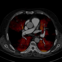
Looking at the 5th dimension of a CT image is old hat. Back in the 1980s there were many installations with rapid kV switching, a dual energy procedure, which were mainly used for bone density measurements. Basically, the 5th dimension is the ability to determine the atomic number as well as density of materials, which facilitates tissue differentiation even when there is the same attenuation.

Nothing has changed regarding the basic principles of the procedure, although nowadays the most up-to-date and fast CT technology enables whole-body scanning – a factor that puts the procedure in the spotlight again, 30 years after its first use – and the interest is considerable. Thus, in the New Horizon session at this year’s ECR, the opportunities from modern dual energy imaging will be aired. Initially, Professor Willi A Kalender, Director at the Institute of Medical Physics at the Friedrich Alexander University in Nürnberg-Erlangen, will provide insights into the basic principles of dual energy CT. This he explained in our European Hospital interview, along with how it could fits in with molecular imaging
‘The dual energy principle, which has gained new relevance since the introduction of the dual source CT in 2005, delivers information about atomic number and density in addition to the “normal”, physical measurement, the linear attenuation co-efficient,’ explained Prof. Kalender. ‘In practice, this can mean the following: both a calcification and a contrast medium have significantly raised CT levels and therefore are almost impossible to distinguish in a normal CT image. However, thanks to the additional information, they can be clearly distinguished – but the additional information is gained at the expense of the signal-to-noise ratio. Therefore, my recommendation is initially to evaluate the normal CT images at the highest resolution and then to retrieve the additional information.’
Why is this relatively old procedure experiencing a second coming?
‘In the 1980s it was only possible to use the procedure for examinations where there were no limiting factors, such as breathing or heartbeat, because of the long examination times. This is why, back then, hardly any examinations other than bone density measurements could be carried out. The speed of modern CT generations now facilitates the examination of the entire tissue and entire body. Therefore, the 5th dimension is indeed opening up very new perspectives. CT contrast perfusion is a good example: In the case of suspected coronary sclerosis or reduced myocardial perfusion, the image shows not just the stenosis but also provides information about haemodynamic significance, i.e. whether there is reduced perfusion of the vessels. Professor Joseph Schöpf, in the USA, is one of the trailblazers in this field, and it was he who also proclaimed the so-called one-stop shopping principle, which states that CT coronary angiography is sufficient in establishing a diagnosis.’
Is the new euphoria justified?
‘Basically, every additional diagnostic benefit also benefits the patient. We don’t yet know in how many cases this additional information is relevant. Some hospitals state that this is the case for about 50% of patients. However, as almost always in the field of medicine, it will take time for a new procedure to become established; this is why we cannot say with any certainty whether or not dual energy will lead to a breakthrough. It is truly a “New Horizon” topic. It is promising, and we have already seen a lot of images, but we need a more extensive, simultaneous evaluation from many sources.’
Where does the 5th dimension fit in molecular imaging?
‘In my view, very well indeed; CT has to escape from Hounsfield’s cage and dual energy finally enables us to offer added value. In the case of cerebral blood flow studies, for example, it allows us to provide estimates on blood volume and mean transit time, in addition to Hounsfield units. In essence, the objective is to extend the range of information provided. Whether or not this should be classed as molecular imaging I don’t know, but the main issue is that it helps the patient.’
02.03.2011


